Osteoporosis
21 Dec 2016
Depression of density and disturbance of structure of a bone tissue is characteristic of an osteoporosis that leads to fractures already at insignificant injuries. Fractures of bodies of vertebrae, a distal part radial and necks femoral bones are most often observed though because of the general fragility of skeleton fractures of other long tubular bones and ribs are frequent.
In the developed countries the osteoporosis at elderly becomes more and more urgent medical problem. It is accepted to distinguish primary and secondary osteoporosis. The secondary osteoporosis develops at general diseases or owing to reception of such medicines as glucocorticoids and Phenytoinum. The most successful way of fight against the secondary osteoporosis is elimination of its reason. However its development is the cornerstone of the same disturbances of updating processes of bone tissue, as at primary osteoporosis; therefore treatment in both cases can be identical.
In 1948 Albright and Reyfenstein came to a conclusion that primary osteoporosis can be a consequence of two independent reasons: depressions of level of estrogens in post menopause and aging. This point of view was supported by Riggs and coworkers. (Riggs et al., 1982) which suggested to distinguish an osteoporosis like I, or post climacteric is loss of spongiform substance of bones at women because of deficiency of estrogens in post menopause and osteoporosis like II, or senile, characterized by loss of both compact, and spongiform osteal substance at men and women because of insufficient efficiency of updating of a bone tissue throughout life, an incomplete delivery and age activation of parathyroid glands. However it isn't proved yet that these states really differ. Besides, the offered scheme doesn't consider that deficiency of mass of a bone tissue can be bound to disturbance of formation of a skeleton during body height. Though at many women loss of mass of a bone tissue after a menopause undoubtedly accelerates, post climacteric osteoporosis, perhaps, it is more correct to consider result of influence of a set of physical and hormonal factors, and also nutrition features.
Structure of bones
Bones are updated with an unequal speed and therefore it makes sense to consider separately bones of an additional skeleton and an axial skeleton. About 80% of all mass of a bone tissue fall to the share of bones of an additional skeleton; they consist mainly of compact substance. Bones of an axial skeleton, for example vertebras, under a thin layer of compact substance contain a lot of spongy substance. Spongy substance consists of closely bound bone plates (trabeculas) reminding bee honeycombs. Between trabeculas there are marrow and fat. For a number of reasons changes of updating of a bone tissue first of all and most deeply affect bones of an axial skeleton. The matter is that processes of updating proceed on the surface of bones, and the surface area of spongy substance is more, than compact. Besides, the marrowy cages predecessors participating in updating of a bone in spongy substance settle down very close to a surface of trabeculas.
Mass of bone tissue
Density of a bone tissue and risk of changes at advanced age depend on the content of mineral substances in a bone tissue by the time of growth termination (that is with the maximum mass of a bone tissue) and on the speed of decrease in this weight further. The greatest gain of mass of a bone tissue (approximately for 60% of maximum) occurs at teenage age that is in days of the largest growth rate. At girls this gain almost comes to an end to 17, and at young men by 20 years. The mass of a bone tissue depends mainly on hereditary factors though also the level of estrogen and androgens in blood, the content of calcium in food and physical activity matter.
Adults have a depression of mass of a bone tissue. X-ray inspections of metacarpal bones (Gam et al., 1966) taped characteristic dynamics of this indicator throughout life: on the third decade of life the gain of mass of a bone tissue stops, up to 50 years it remains to a constant, and then gradually decreases. Such dynamics doesn't depend on a floor and an ethnic origin. It quite precisely reflects change of mass of compact substance, but loss of spongiform substance of some bones begins probably to 50-year age. At women within several years after a menopause loss of mass of a bone tissue accelerates in connection with depression of level of estrogens. You can also like Timusamin.
The mass of a bone tissue at adults depends mainly on physical activity, level of sex hormones and consumption of calcium. All three of these factors are important for its conservation, and a failure of one of them can't be compensated by redundancy of others. For example, at sportswomen with an amenorrhea the mass of a bone tissue decreases, despite intensive exercise stresses (Marcus et al., 1985).
Prophylaxis and treatment of osteoporosis
From the above it is clear in what prophylaxis of an osteoporosis has to consist. Regular exercise stresses of moderate intensity are shown at any age. Children and teenagers have to receive enough calcium with nutrition fully to realize the genetic potential of accumulation of mass of a bone tissue. To people 60 years are more senior special attention should be paid to a delivery: the ration has to contain the increased amount of calcium; also drugs of a calcium and vitamin D are shown. In post menopause as the most effective remedy of conservation of mass of a bone tissue and prophylaxis of fractures serves replacement therapy by estrogens. It is more than that, prophylaxis or treatment of hypogonadism the most important condition of conservation of osteal weight at any age. When keeping of the listed references during all life it is possible to achieve essential depression of risk of fractures.
The medicines applied at an osteoporosis have to or suppress resorption of a bone tissue, or accelerate bone formation. The USA uses only the drugs suppressing resorption now. However resorption of bones and bone formation two parties of one process and therefore the agents suppressing resorption eventually lead to depression and rate of bone formation. Therefore such agents can't provide essential augmentation of density of a bone tissue. The density gain, usually observable in the first years of treatment, occurs due to decrease of units of osteal updating; soon new equilibrium and density of a bone tissue doesn't change any more. To find out whether there are any mechanisms of augmentation of this indicator, whether it is possible process of long clinical tests (not less than 2 years).
In the recent research conducted by scientists from the Spanish and Canadian institutes it was established that additives of melatonin promote strengthening of bones. It opens a possibility of use of such additives as a way of prevention of an osteoporosis at the people having predilection to disease.
Drugs for treatment of osteoporosis
Calcium
The physiological role of calcium and its use at the hypocaltsimic states were considered above. As for its value of osteoporosis prophylactic, it various in the different age periods. At children's and teenage age calcium consumption is a necessary condition of a gain of mass of a bone tissue. In controlled researches it is established that additional reception of calcium promotes augmentation of mass of bone tissue at teenagers (Johnston et al., 1992; Lloid et al., 1993) though it isn't known whether it changes the maximum size of mass of a bone tissue. The increased consumption of a calcium on the third decade of life enlarges a gain of mass of a bone tissue during this period (Recker et al., 1992). Data on expediency of additional reception of calcium at the beginning of the post climacteric period when loss of mass of a bone tissue is bound generally to depression of level of estrogens are contradictory. It was reported about weak influence of calcium on a condition of spongiform substance; however calcium administration of drugs, even against the background of the high content of calcium in nutrition, slows down loss of compact substance (Riis et al., 1987). At elderly the enlarged consumption of calcium slows down updating of a bone tissue, enlarges density of a bone tissue and reduces risk of fractures (Chapuy etal., 1992; Recker etal., 1996; Dawson-Hughes et al., 1997).
At impossibility or unwillingness to enrich a ration with foodstuff with the high content of calcium of the patient can choose one of many nice to the taste and inexpensive drugs of a calcium. There is a huge amount of the drugs containing calcium salts: usually prescribe calcium a carbonate, but there are also Sodium lactatum, gluconat, Natrii phosphas and calcium citrate, and also gvdroksiapatit. Pollution by lead of some consignments of bone meal limits its use as a calcium source. Calcium Citras, apparently, is soaked up better, than other its salts. However all salts of a calcium are soaked up rather well, and for many people the price and taste of drug have larger value, than insignificant differences in efficiency. Usually drugs of a calcium prescribe at the rate of 1000 mg of a calcium a day (such quantity contains, for example, in 1 l of milk). As the usual ration of elderly people contains calcium in number of 500 600 mg/days, the general consumption of calcium at them at the same time increases approximately up to 1500 mg/days. For compensation of losses of endogenic calcium with feces higher doses can be required, but at calcium consumption over 2000 mg/days often arise constipation. Calcium drugs, as a rule, accept during food.
Vitamin D and its analogs
The physiological role of vitamin D and its metabolites, and also their use at hypocalcemia, rachitis and osteomalacy were discussed above. At the boundary or insufficient content of vitamin D in an organism additional reception of its moderate doses (400 800 ME/days) improves a calcium absorption in an intestine, suppresses process of updating of a bone tissue and enlarges density of a bone tissue. Results of two European researches showed that additional reception of vitamin D reduces risk of fractures (Chapuy etal., 1992; Heikinheimoetal., 1992). The purpose of purpose of calcitriol at osteoporosis is not prophylaxis of avitaminosis of D, but suppression of function of parathyroid glands and updating of a bone tissue. Calcitriol and another polar derivative vitamin D is often prescribed in Japan and other countries (Fujita, 1992; Tilyard et al., 1992), however in the USA experience of use of these agents is ambiguous. High doses of calcitriol, apparently, more enlarge density of a bone tissue, but at the same time also the risk of hypercalcuria and hypercalcemia increases. It demands careful observation over the patient and selection of doses. Toxic effect of calcitriol can be weakened, reducing calcium consumption (Gallagner and Goldgar, 1990). Low prevalence of hypercalcuria and hypercalcemia at use of calcitriol in Japan can be bound to rather small consumption of a calcium in this country. Expediency of use of polar derivatives of vitamin D deserves further studying, but their toxicity doesn't allow recommending these agents for broad use yet.
Estrogen
Value of replacement therapy by estrogen in post menopause for preserving mass of a bone tissue and prevention of changes is confirmed with numerous data (Lindsay etal., 1976; Horsmanetal., 1977; Reckeretal., 1977; Hutchinson et al., 1979; Weiss et al., 1980). Researches show that estradiol, affecting osteoblasts, reduces development of SILT-6 and increases development of osteoprotegerin, thereby interfering with mobilization of predecessors of osteoclast (Gi-rasole et al., 1992).
Minimum effective preventive dose of estrogen constitutes 0,625 mg/days of the conjugated estrogen (or an equivalent dose of other medicine). Suppression of updating and preserving mass of a bone tissue are observed both in case of acceptance of estrogen inside, and in case of their application. After cancellation of estrogen decrease in mass of a bone tissue accelerates again therefore treatment shall be long. To women to whom operation on removal of a uterus wasn't performed usually recommend to accept along with replacement therapy by estrogen progestagen (cyclically or constantly). Progestagen, belonging to C21 steroids (for example, medrocsiprogesteron), don't interfere with effect of estrogen on bone tissue. Progestagen with androgenic activity, for example noretisteron, in case of combined use with estrogen increase density of a bone tissue and have additional favorable effect on a skeleton (Christiansen and Riis, 1990). Women with a remote uterus can accept estrogen constantly and without addition of progestagen.
It is better to begin replacement therapy with estrogen right after approach of a menopause when updating of a bone tissue accelerates. However the positive effect of estrogen is observed even at women 65 years are more senior. Many elderly women refuse replacement therapy because of its side effects (in particular, cyclic bleedings). Therefore purpose of such therapy requires individual approach.
Selective modulators of estrogen receptors
A lot of work on receiving the estrogens which are selectively operating on a tissue is carried out. One of such drugs, ralocsifen affects as estrogen a bone tissue and a liver, but doesn't influence a uterus, and affects mammary glands as anti-estrogen (hl. 58). At women in post menopause ralocsifenํ stabilizes and to some extent enlarges density of a bone tissue reducing risk of compression fractures of vertebrae (Delmas et al., 1997; Ettingeret al., 1999). Ralocsifen is applied both to prophylaxis, and to treatment of an osteoporosis.
Calcitonin
The physiological role of calcitonin and its use for treatment of hypercalcemia and Pedzhet's illness were considered above. It considerably suppresses resorption of bone tissue osteoclasts and by that enlarges the mass of a bone tissue at an osteoporosis a little (Gruber et al., 1984; Civi-tellietal., 1988; Mazzuolietal., 1986). Its effect is most expressed at patients with high rate of updating of a bone tissue (Civitelli et al., 1988): the mass of a bone tissue can be enlarged by 10 05%, and then is stabilized. The reason of such effect is decreasing of units number of osteal updating. It is recently shown that use of calcitonin in a dose of 200 ME/days approximately reduces risk of compression fractures of vertebrae at women with an osteoporosis by 40%.
Diphosphonates
Use of these drugs at hypercalcemia and Pedzhet's illness was also discussed above. Diphosphonates were the most effective modern prophylactic and treatments of an osteoporosis. If etidronat sodium can cause osteomalacy, then new diphosphonates suppress a resorption of bone tissue in the doses which aren't oppressing a mineralization of bones. The first of diphosphonates for treatment of osteoporosis was alendronat sodium. Three years' clinical test showed that purpose of alendronat sodium to women in post menopause with low density of a bone tissue and fractures of vertebrae increases density of a bone tissue and reduces risk of repeated fractures (Black et al., 1996). At the women receiving alendronat sodium, the frequency of fractures of vertebrae and other bones (including fractures of a neck of a femur) was about 50% less, than at those who received placebo. In the accompanying research the similar effect of an alendronat of sodium is taped also at women with low density of a bone tissue, but without fractures of vertebrae (Cummings et al., 1998). Alendronat of sodium promotes conservation of density of a bone tissue not only at women in the first years after a menopause (Hosking et al., 1998), but also at men, and also at the patients receiving glucocorticoids (Saag et al., 1998). Now alendronat sodium applies to prophylaxis and treatment of the osteoporosis caused by excess of glucocorticoids, and post climacteric osteoporosis. The recommended preventive dose makes 5 mg/days, and medical 10 mg/days.
Though in clinical tests alendronat sodium in general it was transferred well, sometimes it nevertheless causes esophagitis symptoms. They often manage to be weakened if to wash down a tablet with water in a standing position. If it doesn't help, then before going to bed accept H\K inhibitors '-Atfazy (hl. 37). Less side effects at the same efficiency are observed at reception of alendronat sodium on 40 mg once a week. If, despite it, nevertheless there are expressed esophagitis signs, it is necessary to pass to introduction of a pamidronat sodium, 30 mg by i.v. infusion during 3 h Pamidronat sodium is well transferred each 3 months. At the first introduction there can be pains and small fever, but these phenomena quickly disappear and at the subsequent infusions, as a rule, don't renew. Rizedronat sodium in a dose of 5 mg/days also increases density of a bone tissue and reduces risk of fractures of vertebrae in a post menopause (Harris et al., 1999); soon, apparently, its use at post climacteric osteoporosis will be officially approved. Now one more active diphosphonate is ibandronat sodium is tested.
Thiazide-type diuretics
These drugs don't suppress resorption of a bone tissue immediately, but reduce calcium egestion with urine and reduce loss of a bone tissue at patients with hypercalcuria. Whether thiazidne-type diuretics are capable to have similar effect for lack of hypercalcuria is not clearly though there are data that these drugs reduce risk of fractures of neck femur. Hydro chlorthiazidum in a dose of 25 mg of 1 2 times a day significantly reduces calcium egestion with urine. The doses of thiazidne-type diuretics causing this effect, as a rule, there are less hypotensive doses.
Local irritative agents
Read separate article: The warming ointments
The agents promoting bone formation
Fluorine
Influence of excess of fluorine on a skeleton and value of fluorination of water for prophylaxis of caries was discussed above. Sodium fluoride, stimulating osteoblasts, enlarges the volume of a bone tissue (Baylink et al., 1970; Brianconand Meunier, 1981). Reception of sodium fluoride in a dose of 30 60 mg/days leads to rising of density of spongiform substance at many, though not at everything, sick. According to controlled test (Riggs et al., 1990), sodium fluoride doesn't interfere with emergence of compression fractures of vertebrae, though enlarges density of a bone tissue of vertebrae of lumbar department. At the same time at sodium fluoride reception the risk of fractures of other bones considerably increases. It was noticed, however, that in this test too high doses (75 mg/days) were applied; in other works it was shown that in doses of 30 50 mg/days sodium fluoride reduces risk of any fractures (Mamelle et al., 1988). In later research with use of drug of long action which rather poorly increased concentration of ions of fluorine in a blood depression of risk of fractures was also revealed (et al Cancer., 1994). However other data (Riggs etal., 1990) clearly demonstrate that the gain of mass of a bone tissue doesn't guarantee augmentation of durability of bones and that anyway the therapeutic range of drugs of fluorine is very narrow.
Androgens
Replacement therapy by Testosteron enlarges density of bone tissue at men with a hypogonadism. Androgens promote conservation of density of a bone tissue and at women with an osteoporosis, but use of these agents is interfered by their action. Introduction of Nandrolonum (in the form of decanoat) on 50 mg in oil each 3 weeks enlarges density of a bone tissue of an additional and axial skeleton at women with an osteoporosis, without leading to virilescence. Progestogen having androgenic properties Norethisteronum works with estrogens, promoting augmentation of density of bone tissue at women with osteoporosis (Christiansen and Riis, 1990). However lack of convincing data on risk of fractures doesn't allow drawing a conclusion on expediency of such treatment at women yet. In detail androgens are described in hl. 59.
PTG
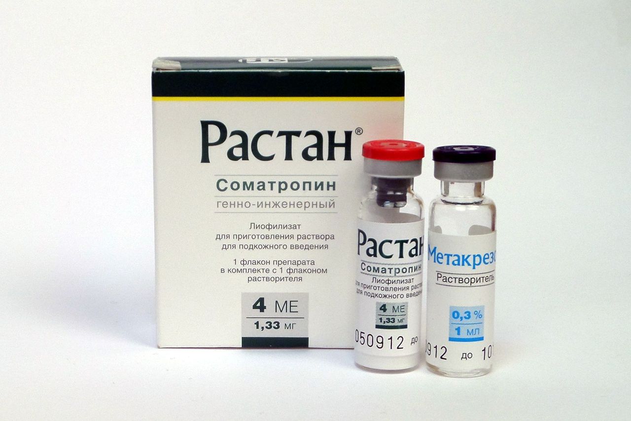
Lesions of bones at the expressed hyper parathyreosis are described above. It is shown, however, that intermittent introduction of PTG has anabolic effect on spongiform substance and therefore in a number of researches influence of PTG on density of a bone tissue at an osteoporosis was studied. According to results of these researches, PTG (134) (a synthetic analog of human PTG) enlarges density of a bone tissue of the axial skeleton (which is generally consisting of spongiform substance), but its action on compact substance was rather negative. At the same time introduction of PTG (134) along with estrogens or synthetic androgens significantly increased density of a bone tissue of an axial skeleton without depression of density of compact substance (Lindsay etal., 1997). PTG considerably enlarges density of a bone tissue at the osteoporosis caused by excess of glucocorticoids (Lane etal., 1998). Now PTG and its analogs pass the 3rd phase of clinical tests.
Influence of protein consumption on development of osteoporosis
Influence of consumption of proteins on loss of a bone tissue was studied in three small researches conducted among women. On the basis of these researches qualitative meta-analysis in which was made "the small advantage of reception of proteins for health of a bone tissue" is noted, but in general data of researches were is regarded as unconvincing from the point of view of influence of reception of proteins on osteal system.
Fenton and colleagues carried out the systematic review and meta-analysis of interrelation between an acid load of a diet, including consumption of proteins, and health of a bone tissue. Unfortunately, data on the considered diets were insufficient, thus quality was estimated by a mark C. Authors noticed that "the carried-out analysis didn't confirm a hypothesis that the acid load of a nutrition can cause an osteoporosis, and also a hypothesis that the "alkaline" diet is capable to prevent an osteoporosis. The high-protein diet significantly doesn't influence calcium level in a bone tissue. At the same time it is represented impossible to define ideal (from the point of view of health of a bone tissue) quantity of the consumed protein".

 Cart
Cart


