Best deal of the week
DR. DOPING

Blog

Logo DR. DOPING

Parathyroidin (parathormone, PTG)

01 Nov 2016

Historical information

Opening of parathyroid glands usually attribute Sendstrema whose article published in 1880 passed almost unnoticed. One decade later these glands were again opened by the Gley which described consequences of their excision together with a thyroid gland. Then the Vassal and Dzhenerali showed that selective excision of parathyroid glands quickly leads to tetany, epileptic seizures and death.

Mack-Kall and Fegglin in 1909 the first described influence of paratireoektomia on concentration of a calcium in plasma. Connection between low concentration of a calcium and symptoms arising at the same time was quickly enough established, and ideas of function of parathyroid glands began to be formed. Active extracts from these glands weakened hypocalcemic tetany at the animals subjected to a paratireoectomia, and at control increased concentration of a calcium in plasma (Berman, 1924; Collip, 1925). These data for the first time allowed to bind clinical disturbances to hyperfunction of parathyroid glands.

While the American and English physiologists studied function of parathyroid glands, the German and Austrian pathologists found a fibrous and cystic ostitis in patients with tumors of these glands. Albright (Albright, 1948) in the fine historical review tracked how these two different ways succeeded to come to the identical conclusions.

Chemical properties

Molecules of paratireoidin at the person, a bull and a pig represent the one-chained polypeptides with a molecular weight about 9500 consisting of 84 amino acids. Their amino-acid sequence is completely deciphered. N-trailer sites of a chain have biological activity. And manifestations of hormonal activity are necessary the amino-acid remains with 1 on 27 for linkng with receptors. The derivatives deprived of the first or second rest though interact with receptors, but are almost not active (Aur-bach, 1988). PTG of a bull and a pig differ with seven amino-acid remains, and the N-trailer site of PTG of the person differs from the respective sites of bull and pork hormones only in four and three remains respectively. Three of these hormones have almost identical biological activity, but differ immunological.

Synthesis, secretion and methods of immunologic analysis

Parathyroidin (parathormone) is synthesized in the form of a polypeptide precursor. In the beginning it is formed so-called preproPTG, consisting of 115 amino acids. At transfer in endoplasmatic retikulum from this polypeptide 25 N-trailer amino-acid remains are split off, and it turns in PTG the Last is had to Golgi's device where from it 6 more amino-acid remains are split off. PTG which is stored in secretory granules is as a result formed and from them it is released in a blood. Normal the most part of PTG is blasted by a proteolysis even before secretion. At hypocalcemia intensity of a proteolysis decreases and cosecretes more parathormone. This mechanism allows to strengthen quickly secretion of hormone without activation of the albuminous synthesis demanding rather big time. At a long hypocalcemia also synthesis of PTG is activated that is followed by a hypertrophy of parathyroid glands. Neither preproPTG nor preproPTG don't get to a blood. Synthesis and processing of a parathyroidin are in detail described, for example, in the review of Kgonenberg of all. (1994).

T1/2 of a mature parathormone in plasma makes 2 — 5 min.; its elimination approximately for 90% is carried out by a liver and kidneys At disintegration of PTG the fragments continuing to circulate in a blood are formed. A part of fragments is formed also in parathyroid glands at PTG proteolysis. Fragments have no biological activity, but react with antibodies to PTG. Nevertheless there are immunologic methods of definition of PTG, rather reliable for the clinical purposes. RIA with use of two types monoclonl the nykh of antibodies (one — to N-trailer, and another — to the S-trailer site of a molecule) has high sensitivity and allows to define the maintenance of mature PTG precisely. This method is used instead of old options of RIA now (Nus-sbaum and Potts, 1994).

Physiology

PTG main function — ensuring constancy of concentration of calcium in blood. Paratireoidin (paratgormon) influences absorption of calcium in intestines, mobilization of calcium from a bone tissue, a calcium ekskretion with urine, a stake, then and milk. Action of PTG on fabric target is mediated by the membrane receptor interfaced to G-protein (hl. 2). Methods of cloning have established the amino-acid sequence of a receptor and an arrangement of seven of his transmembrane domains (Juppner et al., 1991).

Regulation of secretion

The most potent regulator of activity of parathyroid gland is concentration of the ionized calcium in plasma. At its low concentration secretion of PTG increases and if hypocalcemia remains a long time, the hypertrophy and a hyperplasia of parathyroid glands develops. At high concentration secretion of PTG decreases. The researches in vitro showed that low concentration of the ionized calcium in medium for a long time stimulates transport of amino acids in cells of parathyroid glands, synthesis of nucleic acids and protein in them, conducts to augmentation of volume of cytoplasma and intensifying of secretion of PTG. High concentration of a calcium has opposite effect. Thus, a calcium immediately regulates a condition of parathyroid glands, as well as synthesis and secretion of PTG.

Fluctuations of concentration of the ionized calcium in a blood are perceived by the specific membranous receptor located on a surface of cells of parathyroid glands (Brown et al., 1993). Linkng of a calcium with a receptor suppresses secretion of PTG. Hypercalcemia leads to depression of intracellular maintenance of tsAMF and activity of protein, and hypocalcemia activates this enzyme. However communication between these changes and shifts in secretion of PTG is found out not up to the end. Other factors which increase the maintenance of tsAMF in cells of parathyroid glands (for example, p-adrenostimulyatory and Dofaminum) also stimulate secretion of PTG, but in much smaller degree, than hypocalcemia. The active metabolite of vitamin D — calcitriol — suppresses PTG gene expression. Fluctuations of level of Natrii phosphas in a blood in the physiological range, apparently, don't influence secretion of PTG (if, certainly, these fluctuations don't lead to essential changes of concentration of the ionized calcium). At expressed hyper-and hypomagnesiemia secretion of PTG can decrease (Rude et al., 1976).

Thus, concentration of a calcium in a blood is regulated on a feedback mechanism: the regulatory system perceives the level of the ionized calcium in a blood, and in response to it secretion of PTG changes; the last affects various target tissues, enlarging entering of a calcium in a blood.

Influence on bones

Parathyroidin (parathormone) enlarges entering of a calcium from bones in a blood, accelerating process of resorption of a bone tissue during which both organic, and mineral components of intercellular substance are released. PTG cells targets in bones are probably osteoblasts. PTG receptors on osteoclasts are found only in birds. For lack of other cells of PTG doesn't enlarge activity of the osteoclasts applied on a bone surface. Reaction to hormone appears if osteoclasts are cultivated in medium in which there were osteoblasts treated to action PTG before. It testifies to an important role of osteoblasts in stimulation of a resorption of a bone tissue under the influence of PTG (McSheehy and Chambers, 1986; Perry et al., 1987; Takanashi et al., 1988).

Under the influence of PTG precursors of osteoclasts form new units of osteal updating. At long rising of the PTG level there are characteristic histological changes in bones: the number of the centers of a resorption and, respectively, an area of the osteal surface covered with not mineralized organic matrix is enlarged. In this case it indicates not disturbance of a mineralization, and total magnification of the area of bone formation owing to activation of processes of updating of a bone. You can also like Phenazepam.

At PTG incubation with the isolated osteoblasts it usually suppresses their function: decrease formation of collagen I of type, alkaline phosphatase and osteokaltsin. However action of PTG in vivo includes not only influence on separate cells, but also augmentation of total of active osteoblasts as there are new units of osteal updating. As a result the level of an osteokaltsin and activity of an alkaline phosphatase can increase in plasma. Simple model which completely would explain the molecular mechanism of action of PTG on a bone tissue, doesn't exist. The parathyroidin (parathormone) stimulates formation of tsAMF in osteoblasts, but there are data and on a role of an intracellular calcium in oposredon of some effects of this hormone.

Influence on kidneys

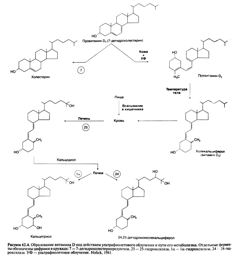
In kidneys of PTG has double effect. First, it strengthens reabsorption of calcium and suppresses a phosphate reabsorption; it leads to increase in concentration in blood of calcium and to decrease — phosphate. Secondly, PTG stimulates transformation of vitamin D into its active form calcitriol (see below). The last contacts receptors in intestines and stimulates calcium absorption that also promotes increase in concentration of calcium in blood. Influence on calcium exchange. PTG strengthens calcium reabsorption in disteel departments of nefron (Agus et al., 1973). Removal of parathyroid glands in case of initially normal concentration of calcium in plasma leads to decrease in its canal reabsorption and by that to increase in its ekskretion with urine. When concentration of this ion in plasma falls lower than 7 mg of % (1,75 mmol/l), its ekskretion decreases: the amount of calcium filtered in balls is so small that it almost reabsorb completely, despite a reduced reabsorb capability of tubules. When entering PTG animal or to the person with canal reabsorption of calcium increases, and the ekskretion decreases. Along with intake of calcium from bones and strengthening of its absorption in intestines it leads to increase in concentration of calcium in blood. When it appears above a regulation, the amount of the filtered calcium begins to exceed possibilities of canal reabsorption (even strengthened under the influence of PTG) and there is a hypercalcium.

Influence on exchange of phosphate

Paratireoidin (paratgormon) increases a kidney ekskretion of phosphate due to decrease in its reabsorption therefore the hypofosfatemia is characteristic of hyperparatireoz.

Action of PTG on kidneys is mediated by tsAMF (Aurbach, 1988). Its activity increases under the influence of PTG is found in cells of cortical substance of kidneys. It leads to strengthening of synthesis of tsAMF which influences canal reabsorption. A part of tsAMF from cages of tubules gets to urine therefore on content of tsAMF in urine judge activity of parathyroid glands and sensitivity of kidneys to PTG. Influence on exchange of other ions. PTG reduces a kidney ekskretion of magnesium, increasing its reabsorption, and strengthens its mobilization from a bone tissue (MacIntyre et al., 1963). Paratireoidin (paratgormon) raises a kidney ekskretion of water, amino acids, citrate, K+, bicarbonate, Na+, SG and SOj ~, but brakes H+ ekskretion. Influence of PTG on a kidney ekskretion of acids and the bases is similar to effect of acetazoleamide, however it isn't mediated by influence on carboangidrasa.

Influence on synthesis of calcitriol

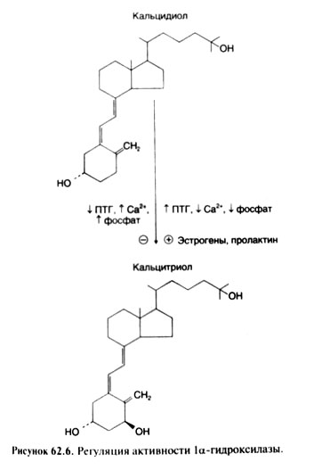
In cells of a renal canaliculus the final stage of activation of vitamin D — its transformation into a hormonal form, that is into calcitriol is carried out (see below). Activity of the la-hydroxylase catalyzing this transformation is regulated by three major factors: Natrii phosphas, PTG and calcium. Depression of concentration of Natrii phosphas in a blood or tissues quickly enlarges formation of calcitriol. A potent stimulator of formation of calcitriol is also PTG whereas hypercalcemia and hyperphosphatemia suppress this process. Thus, when hypocalcemia leads to rising of secretion of PTG, concentration of calcitriol increases in a blood not only because of PTG - dependent depression of concentration of Natrii phosphas, but also owing to direct influence of hormone on activity of a 1a-hydroxylase.

Other effects

Parathyroidin (parathormone) reduces concentration of a calcium in milk and saliva, despite simultaneous rising of its concentration in plasma. Thus, constancy of concentration of a calcium in a blood is provided with also inhibiting influence of PTG on transport of this cation in milk and saliva.

General scheme of a regulation of concentration of a calcium in a blood. Reaction of cells of parathyroid glands even on small depression of concentration of the ionized calcium arises very quickly (within minutes). Correction of short-term fluctuations of concentration of a calcium in a blood is provided with influences of PTG on a calcium reabsorption in kidneys. At longer hypocalcemia in kidneys the 1a-hydroxylase is activated that leads to augmentation of formation of calcitriol; the last stimulates a calcium absorption in an intestine. Besides, a calcium from labile fraction of bones comes to a blood. At a long and serious hypocalcemia processes of updating of bones accelerate (the quantity of units of osteal updating increases); it also leads to a calcium exit in a blood, but already due to depression of mass of a bone tissue.

When concentration of the ionized calcium in plasma is enlarged, secretion of PTG falls and the canalicular reabsorption of a calcium decreases. Besides, depression of the PTG level in a blood promotes Natrii phosphas reabsorption in kidneys. Both depression of the PTG level in itself, and a hyperphosphatemia suppress formation of calcitriol that, in turn, leads to decrease of an absorption of a calcium in an intestine. At last, also process of updating of bones is slowed down.

The scheme of a regulation of concentration of a calcium in a blood is provided. It is visible that 2 hormones (PTG and calcitriol) and 3 effector organs participate in this regulation (kidneys, a GIT and bones). In the provided scheme the role of other hormones isn't considered (for example, a calcitonin), but at the person they, most likely, don't regulate concentration of a calcium in a blood independently, and only modulate activity of the main regulatory system including PTG and vitamin D.

Hypoparathyrosis

This quite infrequent state which is only one of many reasons of hypocalcemia (see above). Most often the hypoparathyrosis arises after operations on thyroid or parathyroid glands. More rare he is is caused by genetic or autoimmune disorders. At pseudohypoparathyroidism biochemical disturbances same as at a hypoparathyrosis, but the PTG level in a blood is increased This state it is caused by resistance of target organs to PTG — usually owing to defect of a receptor, G-protein or adenylatecyclase (Levine, 1999).

Both at hypoparathyrosis, and at pseudohypoparathyroidism the hypocalcemia and symptoms accompanying it are observed. The earliest symptom of hypocalcemia is paresthesia in extremities. The percussion a hammer on nerves causes reduction of the corresponding sceletal muscles (for example, Hvostek's symptom — reduction of face muscles in response to blow by a hammer on a point of an exit of a facial nerve). After this the tetany — tonic cramps of muscles can develop (especially cramps of muscles of brushes and feet and laryngospasm). At last, there are generalized epileptic seizures and other central disturbances. Symptoms from smooth muscle organs are possible — spastic strictures of a ciliary muscle (an accommodation spastic stricture), muscles of an iris, esophagus, intestine, a bladder and bronchi. Changes of an ECG and the expressed tachycardia testify to a heart lesion. Often there are vasospasms of fingers of arms and legs. At chronic hypoparathyrosis changes of tissues of ectodermal parentage meet: hair loss, lamination and fragility of fingernails, defects of an adamantine substance of tooth and cataract; at roentgenography of the head it is possible to find a calcification of basal kernels. Quite often there are mental symptoms: emotional lability, alarm, depression and delirium.

Apply to treatment of hypoparathyrosis and pseudohypoparathyroidism mainly vitamin D (see below). Also calcium additives can be required.

Hyperparatireos

Primary hyperparatireos is caused by hyper secretion of PTG one or several parashitovidny glands. Concentration of calcium in plasma in case of hyperparatireos sometimes remains normal, but is usually increased. Concentration of phosphate is on the lower bound of a regulation or is reduced. Ekskretion of calcium with urine, as a rule, increases, reflecting prevalence of its filtering over canal reabsorption (even strengthened under the influence of PTG). However the calcium ekskretion in case of hyperparatireos nevertheless is less, than in case of hyperkaltsiyemiya of other origin (in case of identical concentration of calcium in plasma). Secondary hyperparatireos develops as compensatory reaction to decrease in level of calcium in blood and isn't followed by a giperkaltsiyemiya. At the same time concentration of phosphate is especially small (if only at the same time there is no renal failure), and activity of alkaline phosphatase in serum is sharply increased.

In case of heavy primary or secondary hyperparatireos special damage of bones is fibrous and cystous osteitis can be observed. However at most of patients with primary hyperparatireos of change of bones are expressed very poorly. Usually they include moderate decrease in general density of a bone tissue at the expense of compact substance; density of spongy substance decreases a little (Bilezikian et al., 1994).

Diagnostics of hyperparatireos was simplified by emergence of methods of RIA allowing to determine the level of mature PTG. The hypercaltsiemia combination to the increased level of mature PTG allows to make the correct diagnosis more than in 90% of cases.

Treatment

Removal of single adenoma of a parathyroid gland (about 80% of cases of hyperparatireos) either removal or subtotal resection of several hyperplasirglands (about 15% of cases of hyperparatireos), carried out by the experienced surgeon, leads to treatment of hyperparatireos. The passing postoperative hypocaltsiemia can be connected with temporary violation of blood supply of the remained ferruterous fabric or with the strengthened calcium absorption by a bone tissue. Fixed hyperparatireos, requiring lifelong treatment by vitamin D and calcic additives — a heavy, but rare complication of transactions on parathyroid glands.

Use of PTG

In last PTG applied to rising of concentration of calcium in plasma, however the same effect with larger safety can be reached administration of calcium and vitamin D. In the list of the resolved FDA of PTG remedies is absent, but use of PTG or its analogs can be approved at an osteoporosis in the near future. It is shown that daily introduction of PTG(1—34) sick with an osteoporosis considerably enlarges the mass of a bone tissue of a backbone (see. "Osteoporosis"). PTG(1—34) can be used for differential diagnostics of hypoparathyrosis and pseudohypoparathyroidism. As resistance of target organs to PTG is characteristic of pseudohypoparathyroidism, administration of drug doesn't raise tsAMF egestion with urine. This assay allows to tap the nature of disturbances at certain patients and their relatives, however the diagnosis is usually established by determination of level of mature PTG in plasma.


Logo DR. DOPING

Androgens

01 Nov 2016

Androgens (Greek ανδρεία (courage, bravery) + Greek γένος (a sort, a knee)) — the general collective name of group of the steroid hormones produced by gonads (testicles at men and ovaries at women) and a cortex of adrenals and having property in certain concentration to cause androgenesis, virilescence of an organism — development of men's secondary sexual characteristics — in both floors.

Women have androgens in concentration, characteristic of men, cause augmentation of the sizes of a clitoris and vulvar lips and rapprochement of vulvar lips (that makes them more similar to a scrotum), a partial atrophy of mammary glands, a uterus and ovaries, the termination of a menses and ovulations, sterility.

Natural androgens

  • Testosterone - an inactive form
  • Digidrotestosteron
  • Degidroepiandrosteron (Dehydroepiandrosterone, DHEA)
  • Androstenedione (Andro)
  • Androstenediol
  • Androsterone

Androgensin bodybuilding

In bodybuilding anabolic steroids are widely applied, however it is necessary to give preference to those medicines which have the smallest androgenic activity, they promote increase in muscle bulk, at the same time cause such side effects less as: the acne, irritability, baldness, seborrhea, a prostate hypertrophy, a masculinization, and the most important, steroids with low activity to a lesser extent cause an atrophy of testicles and after a cycle the natural level of testosterone is restored better and more stoutly. It is connected with the fact that steroids with high androgenic activity contact more androgenic receptors which settle down in a hypophysis and a hypothalamus therefore gonadotrophin level decreases.

Though recently data on androgenic activity are exposed to insinuations from laymans which spread the false information that anabolic and androgenic activity is concepts not applicable to steroid hormones.

Clinical pharmacology

Biosynthesis of androgens

Testosterone — the main androgen at men and, apparently, at women. At men his most part is synthesized in cages of Leydiga. At women testosterone is synthesized in the same way, but in a yellow body and bark of adrenal glands. Predecessors of testosterone androstenedione and dehydroepiandrosterine have weak androgenic activity.

Secretion and transfer by blood

Practically at any age at men more testosterone, than at women is formed, and it explains almost all distinctions between floors. In the I trimester of pregnancy testicles of a fruit begin to emit testosterone (possibly, under the influence of secretable HG placenta) which plays the main role in formation of male genitals. By the beginning of the II trimester its serumal concentration becomes almost same, as in the middle of the pubertal period — about 250 mg % (fig. 59.2) (Dawood and Saxena, 1977; Forest, 1975). By the end of the II trimester she falls, but to childbirth reaches about 250 νγ % again (Forest and Cathiard, 1975; Forest, 1975; Dawood and Saxena, 1977), perhaps, due to stimulation of cages of Leydiga L G, formed in a fruit hypophysis. In the first days of life concentration of testosterone decreases again, then by 2 — 3 months rises to 250 ng % and further falls lower than 50 ng %, remaining at such level prior to the beginning of the pubertal period (Forest, 1975), From 12 — 17 years concentration of testosterone at boys increases much stronger, than at girls, by the end of teenage age reaching 500 — 700 and 30 — 50 ng % respectively. At men high concentration of testosterone provides puberty and further development of secondary sexual characteristics. With age she gradually decreases, bringing, perhaps, various manifestations of aging.

LG, secretable gonadotrophic cells of a hypophysis (hl. 56) — the main stimulator of secretion of testosterone. Perhaps, L G action amplifies in the presence of FSG — the second hormone of these cells. In turn, stimulates secretion of LG gonadropin, formed in a hypothalamus, and brakes testosterone which directly affects gonadotropin cells. Secretion of LG happens pulsewise, the interval between peaks of secretion makes about 2 h, and amplitude of peaks is higher in the morning. Such nature of secretion of LG, apparently, is defined by impulse secretion of gonadoliberin in a hypothalamus. In case of a hypothalamic gipogonadizm impulse introduction of gonadoliberin normalizes L G secretion and testosterone whereas the long infusion of gonadoliberin doesn't help (Crowley et al., 1985).

Testosterone secretion also happens pulsewise and generally in the afternoon. Its concentration is maximum at 8:00 and is minimum at 20:00. With age morning concentration of testosterone decreases (Bremner et al., 1983).

Causes production of testosterone in the yellow body which is formed on the place of a follicle after an ovulation in L G women. However as normal main inhibitors of secretion of LG at women serve the estradiol and progesterone, but not testosterone. In blood about 2% of testosterone are in a free form, 40% strongly are kept by the globulin connecting sex hormones, and other testosterone is unsteadily connected with albumine.

Metabolism

Testosterone has various effect on many fabrics. One of the reasons of such variety — transformation of testosterone into two other steroid hormones, dihydrotestosteron and an estradiol (fig. 59.3). Testosterone causes one effects itself, others are rendered by dihydrotestosteron, the third — an estradiol.

Irreversible restoration of testosterone in dihydrotestosteron is catalyzed by 5a-reductase. Both hormones activate the same androgenovy receptors, but dihydrotestosteron has bigger affinity to receptors (Wilbert et al., 1983) and stronger influences an expression of genes (Deslypere et al., 1992). Turning into dihydrotestosteron in the fabrics containing a 5a-redukgaza, testosterone can make on them additional impact. Two types of 5a-reductase are described: type 1 (mainly in a liver and extregenital sites of skin) and type 11 (in uric ways and genitals at men, and also in skin of genitalia at men and women). Effect of dihydrotestosterone on these fabrics is described below. Do not forget take Cortexin for better results.

Testosterone is inactivated in a liver with formation of an androsteron and etiokholanolon. Dihydrotestosteron turns in androsteron, androstandion and androstandiol.

Physiological effects and mechanisms of action

Effects of testosterone depend on what receptors it affects, and also from fabric and from age of the person. Testosterone renders as androgenic action, contacting androgen receptors directly or after transformation into dihydrotestosteron, and estrogenic, by transformation into estradiol and activation the estrogen of receptors.

Action on androgen receptors. Testosterone and dihydrotestosteron stimulate the same androgen receptors which concern to the group of intracellular receptors including also receptors of steroid hormones, thyroid hormones, calcitriol, retinoid and a number of receptors with unknown ligands. Testosterone and dihydrotestosteron interact with the receptor domain of a receptor that hormone allows - receptorly to a complex by means of the DNA-connecting domain to contact certain genes. The hormone-receptor complex works as a transcription factor, strengthening an expression of these genes (Brinkmann and Trapman, 2000).

Only in recent years there were data on the reasons of a variety of effects of androgens in various fabrics. One of them — higher affinity of dihydrotestosterone to androgenovy receptors in comparison with testosterone (Deslypere et al., 1992; Wilbert et al., 1983). Recently also other mechanism connected with transcription factors (coaktivators and korepressor), specific to various fabrics has been described.

Value of androgen receptor is accurately revealed by consequences of mutations of the gene coding it. As one would expect, the mutations changing primary structure of protein (there is enough replacement of one amino acid in DNA-connecting or receptor domains), cause resistance to testosterone already in the pre-natal period (McPhaul and Griffin, 1999). It leads to violation of a sexual differentiation and a delay of sexual development.

The mutation of other type causes the H-linked bulbospinal amyotrophy (Kennedy's syndrome). At such patients the number of repetitions of TsAG coding a glutamine because of what the polyglutamic site on the N-end of a receptor is extended (Laspada et al is increased., 1991). It reduces sensitivity of a receptor to androgens only a little, but leads to the progressing atrophy of motor-neurons (the mechanism of the last isn't known).

At last, mutations explain development of resistance to anti-androgenic therapy at the spreading prostate cancer. In the beginning the tumor happens hormonal and dependent, at least partly what anti-androgenic therapy is based on. Often in the beginning the tumor will respond to treatment and her sizes decrease, but then stability is developed. At such patients various mutations of a gene of androgen receptor because of what the receptor can be activated by other ligands or in general for lack of a ligand (Visakorpi et al are described., 1995).

Action on estrogen receptors

Transformation into estradiol under the influence of aromatase explains influence of testosterone on bones, and it is possible, and on some other fabrics. In those exceptional cases when in an organism of the man are absent an aromatase (Carani etal., 1997; Morishmaetal., 1995) or estrogenovy receptors (Smith et al., 1994), epiphyseal zones of growth are not closed and tubular bones grow beyond all bounds long; besides, osteoporosis develops. The estradiol eliminates all these violations at insufficiency of an aromatase (Bilezikian et al., 1998), but not at defect the estrogen receptors. There are data that transformation of testosterone into estradiol defines sexual behavior of males of rats, but the similar effect at people is not revealed.

Androgens during the different periods of life

Pre-natal period. Approximately on the 8th week of pregnancy under the influence of HG testicles of a fruit begin to emit testosterone. Local increase in concentration of testosterone in wolffian ducts in internal male genitals causes a differentiation: small egg appendages, seminal ducts and seed bubbles. In laying of genitalia testosterone turns into dihydrotestosteron under the influence of which the penis and a scrotum, and also a prostate gland develop (George and Wilson, 1992). Strengthening of secretion of testosterone by the end of pre-natal development causes growth of a penis.

Newborns. Value of the increased secretion of Testosteron-Depotum in the first months of life isn't known.

Pubertal period. At men this period begins approximately in 12 years with rising of secretion of a gonadoliberin in a hypothalamus. It strengthens development by gonadotrophic cells of FSG and LG which promote body height of testicles. The augmentation of testicles is the first sign of puberty. The strengthened synthesis of Testosteron-Depotum along with action of FSG on Sertoli's cells stimulates development of a wavy seed canaliculus in which spermatozoons are formed. Rising of concentration of Testosteron-Depotum in a blood at the same time influences many tissues, but changes in most of them happen gradually, within several years. Length and thickness of a penis are enlarged, there are scrotum cords, the prostate gland begins to excrete the secret which is a part of a semen. Production of dermal fat increases, the skin becomes more rasping and fat that promotes development of acnes. There are hair in axillary hollows, on a pubis, then on anticnemions and at last on other parts of a body and on a face. Development of a pilosis can take about 10 years, its end speaks about the end of the pubertal period. The weight and force of muscles, especially muscles of a shoulder girdle increase, the hypodermic fat becomes thinner. Tubular bones are quicker extended that leads to the general acceleration of body height, however epiphyseal zones of body height are gradually closed because of what body height is slowed down and as a result stops. Bones are at the same time thickened. As a result of rising of mass of muscles and a bone tissue weight is considerably enlarged. The erythrogenesis amplifies, and concentration of a hemoglobin at men becomes higher, than at boys and women. Cartilages of a larynx are thickened and the voice goes down. Sexual desire develops.

Rising of concentration of Testosteron-Depotum in the pubertal period affects also nervous activity: men are guided in space usually best of all, and their behavior in a number of the relations differs from behavior of women, in particular men are more aggressive.

Mature age. At young and middle age both serumal concentration of testosterone, and characteristic features of the adult man almost don't change. However the androgenetichesky alopetion beginning with emergence of high temples and a hair loss on a darkness gradually develops.

Much more changes in a prostate gland have bigger medical value. First, at all men prostate gland adenoma in a varying degree develops. Sometimes it leads to sdavleniye of an urethra and violation of outflow of urine. Emergence of adenoma is connected with transformation of testosterone into di hydrotestosteron in cells of a prostate gland under the influence of 5a-reductase of type II (Wilson, 1980). One of modern approaches to treatment of adenoma of a prostate gland is based on inhibition of this enzyme (McConnell et al. 1998), as it is described below.

Secondly, the prostate cancer can develop. Though there are no direct proofs of an etiologichesky role of testosterone, the tumor is hormonal and dependent, at least partially and during some time. In this regard at a meta-staziruyushem prostate cancer seek to reduce concentration of testosterone (Huggins and Hodges, 1941; Iversen et al., 1990).

Advanced age. With age serumal concentration of testosterone gradually falls, at the same time concentration of the globulin connecting sex hormones grows. By 80 years the general concentration of testosterone makes about 85%, and concentration of free testosterone — only about 40% of the corresponding concentration in 20 years (Purifoy et al., 1981; Deslypereand Vermeulen, 1984). Such age changes as reduction of working capacity, sexual desire, muscle bulk (Forbes, 1976) and forces can be connected with falling of concentration of testosterone (Murray et al., 1980), and also density of a bone tissue (Riggs etal., 1982). Point out similar changes at a gipogonadizm at young men this dependence (see below).

Deficiency of androgens

Consequences of deficiency of androgens depend on degree of deficiency and age at which it arose. Pre-natal period. In the I trimester of pregnancy deficiency of testosterone at a fruit conducts to an incomplete sexual differentiation. Only changes in testicles can be the cause of deficiency of testosterone (for example, insufficiency of a 17a-hydroxylase): the lack of LG at diseases of a hypophysis or hypothalamus at this stage of development does not result in deficiency of testosterone as in the beginning testosterone secretion by cages of Leydiga is regulated by HG.

For lack of testosterone external female genitals are formed; in less hard cases there is an incomplete virilization and ambiguous genitalia develop, their structure depends on testosterone level. Also the differentiation the volfovykh of channels is broken to semyavynosyashchy canals and seed bubbles, at the same time sekretiruyemy testicles a regression factor the myullerovykh of channels does not allow them to develop in female genitals. Similar changes happen also at normal secretion of testosterone if its activity is reduced owing to defect the androgenovykh of receptors or insufficiency of 5a-reductase. Different defects the androgenovykh of receptors meet. In the most hard cases the receptor is completely inactive, and testicular feminization with development of external female genitals is observed. In medium-weight cases there is their incomplete virilization; in mild cases only the spermatogenesis at adults is broken (McPhaul and Griffin, 1999). Insufficiency of 5a-reductase is followed by an incomplete virilization of genitalia at normal development internal as the last depends on testosterone (Wilson et al., 1993).

Deficiency of Testosteron-Depotum in the III trimester of pregnancy because of illnesses of testicles or deficiency of LG at a fetus leads to two disturbances. First, there is no normal body height of a penis and there are microsinging. It is often observed at boys with L G deficiency caused by disturbance of synthesis of a gonadoliberin. Secondly, testicles don't fall to a scrotum, that is there is a cryptorchism; this state is also quite often observed at L G deficiency.

Pubertal period. If at the boy in the fetal period Testosteron-Depotum is synthesized normally, and its deficiency arises before the pubertal period, then the delay of sexual development is observed. Depending on degree of deficiency the changes of external genitals described above, piloses, the muscle bulk, a voice and behavior are to some extent slowed down. Besides, at normal secretion of STG against the background of Testosteron-Depotum disadvantage of puberty age closing of epiphyseal zones of body height is late that leads to excess elongation of tubular bones and disproportionate body height of arms and legs concerning a trunk because of what the body build becomes eunuchoid. At last, the glandular tissue of mammary glands expands and there is a gynecomastia.

Mature age. If deficiency of Testosteron-Depotum arises after completion of sexual development, there is involution of secondary sexual characteristics, and expression of this process depends on degree and duration of deficiency. In hard cases already in 1 — 2 week depression of sexual desire and working capacity is observed, other symptoms develop more slowly. Depression of muscle bulk and force on average happens in several months, but for emergence of noticeable changes in certain patients years are required.

Within several months there is an essential depression of a hematocrit and concentration of a hemoglobin. The two-photon x-ray absorbtsiometriya is capable to find depression of density of a bone tissue in 2 years though the risk of fractures increases only in many years. Decrease of a pilosis on men's type also happens gradually, for many years. Deficiency of androgens at women reduces a pubic and/1 axillary pilosis, but it becomes noticeable only in several years. At women androgens, perhaps, perform also other important functions lost in their absence (especially at the combined deficiency of ovarian and adrenal androgens against the background of an apituitarism). Testosteron-Depotum drugs which can maintain physiological concentration of this hormone at women are now developed. Use of these drugs will allow to learn how replacement therapy by Testosteron-Depotum at deficiency of androgens at women is capable to raise sexual desire, weight and force of muscles, density of a bone tissue and working capacity.

Medicines of androgens

At intake testosterone is inefficient: he is well soaked up, but quickly inactivated in a liver. Therefore for maintenance of normal serumal concentration of testosterone medicine should be accepted too often and in too high doses. Respectively, the majority of medicines of androgens have the structure interfering their fast destruction in a liver. Besides, search of the medicines possessing more selective action is conducted.

Medicines of androgens

Medicines of androgens for skin application

Other opportunity to avoid an inactivation of the entered testosterone is use of a plaster from which not changed hormone slowly is soaked up through skin. Daily gluing of a plaster allows to reduce fluctuations of concentration of testosterone in comparison with introduction of his air in oil. The first such plasters were pasted on scrotum skin (Findlay et al., 1989). Here skin so thin that testosterone is soaked up in enough and without addition of the substances facilitating absorption. Then the plasters containing such substances have been developed thanks to what these plasters could be pasted on other sites of skin (Yu et al., 1997; Dobs et al., 1999). Testosterone medicine in the form of aqueous-alcoholic gel is recently created (Wang et al., 2000). All these medicines put 1 time a day that provides normal concentration of testosterone at the majority sick gipogenitalism.

Search of selective androgens

Alkylandrogens

Half a century works on synthesis of analogs of testosterone with prevalence of anabolic activity over androgenic were conducted back. It seemed, a number of substances had such properties as at rats they exerted a greater influence on the muscle lifting back pass than on forward department of a prostate gland (Hershberger and Meyer, 1953). These substances were called anabolic steroids, most of them was 17 alkylandrogens. However any of them didn't show desirable selectivity at people. Nevertheless anabolic steroids are widely applied by athletes as dope (see below). Feature of other alkylandrogen, 7a-methyl-19-nortestosterona — resistance to effect of 5a-reductase (Kumar et al., 1992). The selective modulators androgenovykh of receptors. Creation of the selective modulators estrogen receptors (tamoxifen, raloksifen) activating estrogenovy receptors in one fabrics and blocking in others stimulated development of similar modulators the androgen receptors

However, selectivity of raloksifen, apparently, is connected with big affinity to one isoform the estrogen receptors prevailing in bones and in a myocardium and smaller affinity to other form characteristic of a mammary gland and endometrium. As only one form androgen receptors is found, selective action on them has to be based on fabric specificity of coactivators and corepressor — the proteins regulating influence of intracellular receptors on a transcription of genes targets (Moilanen et al., 1999). The group of quinolines with selective androgenic activity is received (Zhi et al., 1999).

Application of androgens

The obvious indication to purpose of androgens — deficit of testosterone (gipogonadizm) at men. Androgens apply also in other cases; possibly, new indications to their appointment in the future will appear.

Men hypogenitalism. In case of deficit of testosterone appoint any of testosterone medicines described above for skin application or testosterone air. At teenagers and elderly it is necessary to watch efficiency and safety of treatment especially carefully.

efficiency evaluation

The treatment purpose — maintenance of serumal concentration of testosterone as is possible closer to normal therefore measurement of this concentration serves as the main evaluation method of efficiency of treatment. Time of measurement depends on medicine: if testosterone medicines for skin application are used, concentration can be determined in any day and at any time. At the same time it is necessary to consider that concentration reaches a maximum in 2 — 4 h after gluing of a plaster for scrotum skin (Findlay et al., 1987) or a plaster Testoderm for the ekstragenitalnykh of sites of skin (Yu etal., 1997) and in 6 — 9 h when using a plaster Androderm for the ekstragenitalnykh of sites of skin (Dobs et al., 1999), and the minimum concentration before gluing of the following plaster makes 60 — 70% maximum (Findlay et al., 1987). When using gel serumal concentration of testosterone within a day changes a little, but stationary concentration of medicine is sometimes established only in a month after an initiation of treatment. When using testosterone of an enantat or tsipionat, entered each 2 weeks, concentration of testosterone is measured in the middle of this interval. The measured concentration of testosterone shan't differ from normal, otherwise the mode of introduction is changed. If deficit of testosterone is caused by defeat of testicles (in this case concentration of LG is increased), normalization of concentration of LG within 2 months after the beginning of replacement therapy testifies to efficiency of treatment (Snyder and Lawrence, 1980; Findlay etal., 1989).

Normalization of serumal concentration of testosterone at men with hypogonadism leads to completion of development of secondary sexual characteristics and their maintenance. Within several weeks at such men sexual desire and working capacity have to raise (Davidson et al., 1979). In several months the muscle bulk and force increase and the mass of fatty tissue decreases (Katsnelson et al., 1996). Density of a bone tissue reaches a maximum within 2 years (Snyder et al., 2000).

Side effects of Testosteron

Testosteron-Depotum drugs for cutaneous use and Aethers of Testosteron don't possess any action other than action of endogenic Testosteron (if the therapeutic dose isn't exceeded). However when performing replacement therapy there can be undesirable effects. Some of them appear soon after an initiation of treatment, others — usually only in several years. Rising of concentration of Testosteron from the level of prepubertatny or the middle of the pubertal period to the level characteristic of the adult man, can conduct to such phenomena inherent to the pubertal period as an acne, a gynecomastia and aggressive sexual behavior. Physiological quantities of Testosteron don't influence a blood lipid profile. Replacement therapy sometimes has undesirable effect at associated diseases. So, stimulation of erythrogenesis leads to normalization of hematocrit at the man healthy for the rest, however at predilection to hyperglobulia (for example, against the background of HOZL) the hematocrit can exceed norm. In a similar way the small delay of sodium and water won't affect the healthy person, but will aggravate a heart failure. At Testosteron overdosage the hyperglobulia and, occasionally, a water delay with edemas develop also for lack of predisposition to these states. When normal concentration of Testosteron is maintained for many years (due to its endogenic secretion or replacement therapy), 40 years are aged more senior the risk of such hormonal and dependent states as adenoma and a prostate cancer increases.

Testosteron derivatives 17 alkylandrogens possess a side effect on a liver, sometimes causing a cholestasia and, occasionally, οελθξη a liver (formation of the lacunas filled with a blood). Separate cases of pechenochnokletochny cancer are described, but the etiological role of these drugs is doubtful. Besides, 17 alkylandrogens, especially in high doses, reduce the LPVP level.

Hypogonadism in the pubertal period. If the hypogonadism is observed at boys, then in the pubertal period prescribe it Testosteron drugs, proceeding from the principles stated above. It is important to remember that Testosteron accelerates closing of epiphyseal zones of body height therefore at first it causes rapid growth, but then body height finally stops. Respectively, it is necessary to consider body height of the boy and the STG level at it. With a low growth and deficiency of STG at first prescribe somatropin, and already then treat hypogonadism.

Aging at men. According to preliminary data, replacement therapy by Testosteron at men with age depression of concentration of this hormone increases density of a bone tissue and body weight and reduces the mass of fatty tissue (Snyderet ah, 1999a, b). However it isn't clear whether body height of adenoma of a prostate causes such treatment, and whether the risk of the prostate cancer which is shown clinically is enlarged. Hypogonadism at women. It isn't proved yet that purpose of Testosteron to women with the lowered serumal concentration of Testosteron promotes body height of sexual desire, working capacity, muscle bulk and force, and also density of a bone tissue.

Dope. For achievement of the best results some athletes resort to dope, including use androgens. Usually these drugs accept secretly therefore their action is studied worse, than effect of the drugs prescribed at treatment of hypogonadism. Drugs. Athletes used almost all androgens applied in medicine and veterinary science as dope. It began more than 20 years ago, and initially preference was given to anabolic steroids — 17 alkylandrogens and other substances which were allegedly possessing mainly anabolic action in comparison with Testosteron. As such bonds can be easily taped by the controlling larger distribution was gained by HG and Aethers of Testosteron increasing serumal concentration of Testosteron-Depotum. In recent years use of precursors of Testosteron (androstendion and degidroepiandrosteron) which aren't entering lists of banned drugs was enlarged. Efficiency. The majority of the tests devoted to influence of Testosteron on an animal force were uncontrollable. In one double blind test 43 persons (all men) were divided into 4 groups: in the first two groups examinees carried out power exercises against the background of introduction of Testosteroni oenanthas in a dose of 600 mg once a week (more than 6 times higher, than at replacement therapy) or placebo, in the third and fourth groups examinees received the same drugs for lack of exercises. Testosteron enlarged body weight and an animal force, and exercises gave additional effect (Bhasin et al., 1997).

In other double blind test androstendion (on 100 mg 3 times a day during 8 weeks) didn't lead to increase in muscular force in comparison with placebo. However, it isn't surprising as average concentration of testosterone also didn't increase (King et al., 1999).

Side effects of androgens

At reception of therapeutic doses of any androgen some side effects arise always; emergence of other effects depends on medicine or additional conditions. All androgens in high doses suppress secretion of LG and FSG, oppressing functions of testicles: production of endogenous testosterone and spermatozoa falls that ability to fertilization. Reception of androgens for many years can lead to reduction of testicles. Function of testicles is usually restored in several months after medicine cancellation, but sometimes more time for this purpose is required. High doses of all androgens cause erytrocytosis (Drinka et al., 1995);

Androgens capable to turn into estrogen (first of all testosterone), in high doses cause ginecomastia. Medicines with the modified ring and, not exposed to aromatization, for example dihydrotestosteron, are deprived of this action.

Hepatotoxic action is peculiar only to 17 alkylandrogens (see above). Besides, in high doses they are more often than other androgens break lipidic exchange, reducing the LPVP level and increasing the LPNP level. There are unconfirmed messages on such side effects as mental disorders and the sudden death from IBS which is presumably connected with change of a lipidic profile and increase in coagulability of blood.

Some side effects of androgens are especially noticeable at women and children — character of pilosis peculiar to men, high temples and eels. At boys the penis increases, women have a clitoris. Boys and girls stop growing because of premature closing the epiphyseal zones of growth. Contraception at men. The contraceptives for men containing androgens are developed (including in combination with other medicines). Their action is based on suppression of secretion of LG in a hypophysis and the subsequent reduction of synthesis of endogenous testosterone. Normal concentration of testosterone in testicles approximately by 100 times exceeds that in blood. High concentration of testosterone is necessary for a spermatogenesis, her decrease sharply slows down this process. However, in the first attempts of use of testosterone suppression of a spermatogenesis required twice a big dose of testosterone enantat, than for replacement therapy, and all the same it wasn't possible to suppress completely a spermatogenesis at all men (WHO Task Force for the Regulation of Male Fertility, 1996). In other early tests the antagonists of gonadoliberin suppressing secretion of LG in combination with replacement therapy by testosterone applied (Pavlou et al., 1991). However such combination isn't suitable for broad application as the available antagonists of gonadoliberin it is necessary to enter in the form of the daily injections causing emission of a histamine. The combination of progestagen to physiological doses of testosterone for suppression of secretion of L G and a spermatogenesis at preservation of normal serumal concentration of testosterone is more perspective (Bebb et al., 1996). Now pass testosterone test undecanoat for injections, providing rather stable serumal concentration of testosterone within a month (Zhang et al., 1999), and 7a-methyl-19-nortestosteron, the synthetic androgen which isn't restored by 5a-reductase and therefore not acting on a prostate gland (Cummings et al., 1998).

Exhaustion. Anabolic activity of testosterone was tried to be used for treatment of an atrophy of muscles and exhaustion, but in most cases this method was inefficient. The exception makes treatment of the atrophy of muscles at AIDS which is followed by gipogonadism. At patients with AIDS of men with an atrophy of muscles and the lowered serumal concentration of testosterone such treatment increases the muscle bulk and force (Bhasin et al., 2000).

Quincke's edema. Continuous treatment by androgens prevents Quincke's edema attacks. Illness is bound to heriditary failure of an inhibitor of C1 esterase or development of antibodies to it (Cicardi et al., 1998). 17 alkylandrogens (stanosolol and danasol) stimulate synthesis of an inhibitor of C1 esterase in a liver. Unfortunately, they cause virilescence in women. Because of virilescence and premature closing of epiphyseal zones of body height androgens don't use for prophylaxis of Quincke's edema at children, but sometimes prescribe them at attacks. Illnesses of a blood. Before emergence of erythropoetin androgens applied to stimulation of an erythrogenesis at anemias of various etiology. Androgens (in particular, danasol) still sometimes prescribe at the hemolitic anemia and an idiopathic Werlhof's disease steady against standard treatment.


Logo DR. DOPING

Fluorine

01 Nov 2016

Compounds of fluorine are important for the doctor, first, because of their toxicity, secondly — as means of strengthening of teeth and bones. These connections are widespread in the nature, but their content in the soil of various regions of the world not equally. In case of combustion of brown coals and production of superphosphate, aluminum, steel, lead, copper and nickel fluorine is emitted in the atmosphere. In a human body it arrives generally with products of plant origin and water.

Absorption, distribution and elimination

Compounds of fluorine arrive in an organism mainly through a GIT, and also through lungs and skin. Extent of their absorption depends on solubility. Rather well soluble connections, such as sodium fluoride, are soaked up almost completely whereas badly soluble, such as cryolitas (NajAlF6) and the fluorides which are present at bone meal (ftorapatit), are almost not soaked up. The second major way of intake of fluorine to an organism — through lungs. Inhalation of compounds of fluorine with dust and gases — a basic reason of poisoning with fluorine on production.

Fluorine contains in all bodies and fabrics, but accumulates in bones, teeth, a thyroid gland, an aorta and probably in kidneys. Most of all fluorine contains in bones and teeth, and its content depends not only on consumption, but also on age. Fluorine inventories reflect the speed of its updating in a bone tissue: in his growing bone it is more, than in bones of mature animals.

Fluorine is removed generally by kidneys, but its small amounts are found in sweat, milk and allocations of intestinal crypts. In case of strong sweating nearly a half of all removed fluorine can be lost. In kidney tubules about 90% of the filtered fluorine reabsorb.

Pharmacological effect

Effect of fluorine (except for probably action on bones and teeth) is toxic. Ions of fluorine inhibit a series of ferment systems, slow down tissue respiration and an anaerobic glycolysis. In vitro fluorine ions, binding Sa, interferes with a blood coagulation. At the same time they oppress glycolysis in erythrocytes therefore they are used at capture of a blood for definition of concentration of a glucose. Add some Nootropil to your daily training.

Ions of fluorine stimulate division of bone-forming sells and bone formation (Baylink et al., 1970). For this reason its bonds try to apply at an osteoporosis. At many, but not at all patients receiving fluorine salts significant increase in mass of spongiform substance of bones became perceptible; compact substance reacts worse. It is necessary to find out whether durability of bones is enlarged at the same time and whether the risk of fractures decreases. Radioactive isotope F is used for scintigraphy of bones (Jones et al., 1973).

Acute toxic exposure

Such poisoning is not a rare thing. Usually it arises owing to casual hit in an organism of fluorinated insecticides or agents against rodents.

The first symptoms (hypersalivation, nausea, abdominal pain, vomiting and diarrhea) are bound to local action of bonds of fluorine on mucous a GIT. Systemic symptoms are various and serious: the nervous exaltation (caused, apparently, by Ca-binding action of ions of fluorine), hypocalcemia and hypoglycemia. The ABP decreases that it is possible to explain both with oppression of the vasomotor center, and direct cardiotoxic action. Respiration becomes frequent in the beginning, but then is oppressed. Death usually comes from respiratory standstill or a heart failure. The lethal dose of sodium fluoride for the person makes about 5 g, though considerably fluctuates. Treatment comes down to i.v. introduction of normal saline solution with a glucose and to a gastric lavage lime water (0,15% calcium hydroxide solution) or solutions of other salts of a calcium for binding of ions of fluorine. At a tetany enter a calcium gluconat i.v. By means of intensive infusional care support a high diuresis.

Chronic poisoning

This poisoning is called flyuorosis. At the person it is shown generally by osteosclerosis and spotty defeat of enamel. Increase in density of a bone tissue owing to activation of osteoblasts and replacement of a hydroxyapatite with more dense ftorapatit is characteristic of osteosclerosis. At a X-ray analysis damage of bones of different degree comes to light: from hardly distinguishable changes to the expressed thickening of long tubular bones, multiple ekzostoz with calcification of sheaves, sinews and sites of an attachment of muscles. In the most hard cases of the patient loses working capacity and becomes the cripple.

Spotty defeat of enamel — the well-known pathology for the first time described more than 60 years ago. Opaque pretty spots of irregular shape appear in mild cases on tooth enamel. In hard cases on the surface of teeth the separate or merging dark brown or black deepenings are visible. Defeat arises because of violation of ability of cells of tooth to production of enamel. Such violation can have the different reasons, and excess intake of fluorine in an organism — only one of them.

Spotty defeat of enamel — a disease of the formed teeth, excess of fluorine doesn't affect the cut-through teeth any more. It is one of the first visible signs of a flyuoroz at the child. Constant water consumption with concentration of fluorine can lead about 1 mg/l only to very minor changes of enamel at 10% of children; at concentration of fluorine of 4 — 6 mg/l damage of teeth, and much more expressed is noted almost at 100% of children.

Earlier in some regions of the world, for example to Pompeii (Italy) or near the mountain Payks-pik (the State of Colorado, the USA) where water contains a large amount of fluorine, the expressed defeat of tooth enamel met very often. In water of some droughty areas in the southwest of the USA fluorine is present at very high concentration, and damage of bones is widespread among the cattle which is grazed there. Now the federal legislation of the USA demands decrease in level of fluorine in drinking water of such areas or uses of other sources of water supply. Constant water consumption with concentration of fluorine of 4 mg/l leads to reduction of mass of compact substance of a bone and acceleration of a resorption of bone tissue over time (Sowers et al., 1991).

Fluorine and caries

Importance of maintenance of normal concentration of fluorine in drinking water received unexpected confirmation when it turned out that at the children born in Boksayte (the State of Arkansas, the USA) in after transition to water consumption without fluorine caries became occurs much to a bowl, than at those who continued to use the water containing fluorine. The large-scale researches conducted by Service of public health care of the USA convincingly showed that addition to water of a small amount of fluorine (1 mg/l) — the safe and effective measure providing essential depression of prevalence of caries of second teeth.

Consumption of the fluorinated water is useful for children of any age, but especially to those at whom second teeth didn't appear yet. Local putting fluorinated solutions by the stomatologist especially effectively if it is begun right after a teething; it reduces risk of caries by 30 — 40%. Aged up to 12 years if drinking water contains fluorine less than 0,7 mg/l, the use of fluorinated nutritional supplements is shown to children. Data on influence of fluorinated toothpastes are contradictory.

Inclusion of fluorine in tooth fabric gives to an outer layer of enamel big hardness and does it to steadier against demineralization. At the same time fluorine, forcing out, apparently, other anions (a hydroxyl and citrate), takes their place on a surface of crystals of a hydroxyapatite. The mechanism of preventive effect of fluorine concerning caries isn't absolutely clear. Efficiency of fluorine after final forming of second teeth (as a rule, after 14 years) remains unproven.

Often add fluoride of sodium or tin to toothpastes. Fluoride of sodium is present also at many means for local application — tablets, drops, rinsings and gels. Insecticides usually contain sodium fluoride, fluosilicate sodium (Na2SiF6) and cryolite.

From the very beginning voices against addition of fluorine in drinking water were distributed. In certain cases it was net political rhetoric, but also concerns expressed that the fluorinated water negatively will affect health. According to results of the careful researches organized by National institute of cancer and Service of public health care of the USA, death rate indicators from oncological diseases and general death rate in the national groups consuming the fluorinated and not fluorinated water significantly don't differ (Hoover et al., 1976; Erickson, 1978).


Logo DR. DOPING

Diphosphonates

01 Nov 2016

Diphosphonates are called group of bonds in which two phosphonew groups are attached to one carbon atom.

Diphosphonates are called group of bonds in which two phosphonew groups

At addition to solutions to Natrii phosphas calcium suspensions these bonds slow down education and dissolution of crystals of hydroxyapatite. The first diphosphonate offered for a clinical use was etidronat sodium, is stronger than other drugs of this group suppressing a mineralization of bones. Further it became clear that suppression of a mineralization is undesirable as it leads to osteomalacy over time. Therefore the diphosphonates of the second and third generation less suppressing a mineralization of bones were framed. Use of diphosphonates is caused by their ability to oppress a resorption of a bone tissue. The mechanism of such effect isn't absolutely clear. Believe that diphosphonates join in an osteal matrix and are absorbed by osteoclasts at resorption. Diphosphonates influence osteoclasts in at least two different ways. Etidronat of sodium, clodronat sodium and tituldronat sodium turn into an adenosine-5 '-f, at - a dichloromethylene) triphosphate (an ATP analog) which collects in cells, breaking their function and reducing viability (Frith etal., 1996). On the contrary, highly active aminodiphosphonates (for example, alendronat sodium and ibandronat sodium) aren't metabolized, and immediately inhibit many stages of transformation of a mevalonat into a cholesterin and izoprenoidny lipids (for example, a geranyl-geranildifosfat). These lipids are necessary for a prenilirovaniye of many proteins playing an important role as osteoclasts (Luckman et al., 1998). One of the Best Nooropic drug is Cerebrolysin.

Now in the USA several diphosphonates are issued. Etidronat of sodium use in case of Pedzhet's disease; it is also entered in/in in case of a giperkaltsiyemiya. As edidronat sodium is the unique diphosphonate suppressing a mineralization it will probably be soon replaced with newer medicines of this group. Pamidronat of sodium (fig. 62.7) is approved as remedy for giperkaltsiyemia, but it turned out that it is effective in case of a number of diseases of bones. In the USA pamidronat sodium is issued only for parenteral application. In case of giperkaltsiyemia enter 60 — 90 mg of pamidronat of sodium in the way to infusions during 4 — 24 h. A number of new diphosphonates are allowed for use in case of Pedzhet's disease. Treat with tiludronat sodium, alendronat sodium and risedronat sodium. All diphosphonates are very badly soaked up in intestines. Therefore in them it is necessary to accept on an empty stomach and, at least, in 30 min. prior to a breakfast, washing down with a complete glass of water.

Use:

Pedzhet's illness

This lesion of a skeleton at which the centers of the broken updating of a bone tissue appear. Pathological "variegation" of bone formation is characteristic of affected areas; in them there is a set of abnormal multinuclear osteoclasts. The structure of bones is broken, they are thickened. Changes of bones sometimes are followed by secondary symptoms, such as pain, a hearing loss, a prelum of a spinal cord and a heart failure with high cordial emission. An infrequent, but lethal complication of illness of Pedzhet is osteosarcoma. Diphosphonates and calcitonin normalize biochemical indicators of updating of a bone tissue: reduce activity of an alkaline phosphatase in Serum and hydroxyproline egestion with urine. Usually treatment is begun with purpose of diphosphonates of 1 times a day within 6 months. At most of patients at the same time ostealgias weaken in several weeks. Sometimes treatment causes long remission. At renewal of symptoms repeated courses of treatment can help. Introduction of high doses etbdronata sodium (10 — 20 mg/kg/days) or its continuous use is accompanied more than 6 months by great risk of osteomalacy. Focal osteomalacy sometimes develops when using and smaller doses of an etidronat of sodium (5 — 7.5 mg/kg/days). When using other diphosphonates or calcitonin of disturbance of a mineralization weren't observed.

Each patient needs to pick up the treatment method which is most suitable for him. The benefit of diphosphonates is that they can be accepted inside, they are rather inexpensive, neimmunogenna also cause side effects less than calcitonin. However calcitonin — very well-tried remedy capable besides to stop bone pains. The bigger effect in case of treatment of a disease of Pedzhet can achieve, apparently, by a combination of diphosphonates and calcitonin (O’ Donoghue and Hosking, 1987). In complex cases sometimes use plicamicin. However it is very toxic and can't be recommended as regular remedy for a disease of Pedzhet.

Hypercalcemia

Etidronat of sodium and pamidronat sodium with success were applied at paraneoplastic hypercalcemia. Etidronat of sodium used also at an ossification of soft tissues or an ossifying miositis counting on ability of this drug to suppress a mineralization. Results, however, were not too impressive.

Postclimacteric osteoporosis

Now diphosphonates draw great attention as agents for treatment of an osteoporosis. Recent clinical tests taped their ability to enlarge density of a bone tissue and to reduce risk of fractures at this disease.


Logo DR. DOPING

Calcitonin

01 Nov 2016

Historical information

In 1962 Kopp discovered the hormone reducing calcium level in a blood, having called calcitonin (Soar, 1964). It was shown that perfusion parathyroid and thyroid glands of dogs a blood with high concentration of a calcium causes fast short-term hypocalcemic effect, and this effect develops earlier, than after a full paratireoektomia. Kopp concluded that parathyroid glands in response to hypercalcemia cosecrete a calcitonin and thus normalize concentration of calcium in plasma. Munson and others (Hirsch et al., 1963) paid attention that the paratireoektomia by electrocoagulation causes in rats more expressed hypocalcemia, than excision both thyroid, and parathyroid glands together that indicated presence of a gipokaltsiyemichesky factor at a thyroid gland. Having shown that extracts of a thyroid gland really cause a hypocalcemia, these authors called a required factor tirokaltsitoniny. Now it is known that both factors represent the same substance and that it is really produced in a thyroid gland. However the name a calcitonin was assigned to this hormone.

Calcitonin is developed and cosecretes the parafollicular S-cells of a thyroid gland developing in an embryogenesis from an ectoderm of a nervous crest. At the vertebrata who aren't belonging to a class of mammals, the calcitonin is present at the ultimobranchial little bodies located out of a thyroid gland. At the person of the S-cell are localized not only in a thyroid gland, but also in parathyroid glands, and also in a thymus.

Structurally functional dependence

Calcitonin is a one-chained peptide from 32 amino acids. Eight of them, including S-trailer prolineamidum PP and Cysteinums in provisions 1 and 7 (forming a disulfide ponticulus), are conservative and necessary for implication of biological activity of hormone. The amino-acid remains of the middle of a molecule (in provisions 10 — 27) are variable and, apparently, influence activity and duration of action of a calcitonin. The calcitonins allocated from ultimobranchial little bodies of a salmon and eel have larger activity of in vivo and in vitro, than hormone from thyroid glands of mammals, and differ from calcitonin of the person in the amino-acid remains respectively in provisions 13 and 16. The calcitonin of a salmon renders larger medical effect, than hormone of the person, partly because possesses larger T1/2.

At the person the calcitonin is formed of the pro-peptide consisting of 135 amino-acid remains; two more peptides which biological role isn't known are formed of the same pro-peptide. The gene of calcitonin contains six exons. Calcitonin is coded by the 4th exon. In S-cells the splaysing occurs in such a way that in final MRNK only the first 4 exons are presented. In nervous cells the sequence corresponding to the 4th exon is cut out, and in final MRNK exons 1—3, 5 and 6 are presented. After broadcasting and splitting of a pro-peptide the mature peptide consisting of 37 amino-acid remains — a kaltsitoninopodobny peptide is formed. At some animals action of this peptide is partly similar to action of a calcitonin, but at others the kaltsitoninopodobny peptide shows PTG-like activity and interacts with the receptors different from calcitonin receptors. As S-cells of a thyroid gland almost don't form this peptide, it hardly takes part in a calcium exchange regulation. The Kaltsitoninopodobny peptide and its receptors are widely presented to a CNS where it probably performs function of a mediator. This peptide is found in many diaxones of spinal ganglions and possesses the expressed vasodilating action. In more detail about a structure and synthesis of a calcitonin see in the review of MacIntyre etal. (1987).

At plasma there are various forms of calcitonin, including high-molecular units and cross connected molecules. It complicates development of reliable immunological methods of his definition. Methods of definition of intact monomeric peptide are offered (Body and Heath, 1983). Secretion regulation. Synthesis and secretion of calcitonin are regulated by concentration of the ionized calcium in plasma. At high concentration secretion of calcitonin increases, and at low — decreases or stops. At the person normal concentration of calcitonin doesn't reach and 10 pg/ml (Body and Heath, 1983). Women have its average concentration and reaction to secretion stimulators (pentagastrin and calcium) less, than at men. Calcitonin T1/2 is equal in blood to about 10 min.

Secretion of a calcitonin is stimulated with many factors, including catecholamines, glucagon, gastrin and kgolecistokinin, but their role in physiological regulation of secretion of calcitonin is doubtful. It isn't known even whether calcitonin plays an essential role in exchange of calcium at the person. Removal of a thyroid gland after which the calcitonin from blood disappears doesn't affect neither calcium exchange, nor the mass of a bone tissue. At medullary cancer of a thyroid gland in plasma, urine and tumoral tissue of patients of concentration of calcitonin are very high (50 — 5000 times higher than norm). This tumor develops from parafollicular cages; all her manifestations are connected a lot of calcitonin. For diagnostics carry usually out test with pentagastriny or calcium: compare plasma calcitonin levels before infusion of pentagastrin or calcium of gluconate (Wells et al., 1978). As one of forms of medullary cancer of thyroid gland is inherited as a prepotent sign (at a multiple endocrine neoplasia like II), the corresponding researches need to be conducted regularly and at the patient's relatives, since the early childhood (Tashjian etal., 1974). In most cases a multiple endocrine neoplasia like II RET protooncogene mutation is found; it allows to hope that to replace tests with pentagastriny and calcium the genodiagnostika will come (Donis-Keller etal., 1993; Carlson et al., 1994).

Action mechanism

Calcitonin reduces concentration of calcium and phosphate in blood mainly due to the direct oppressing action on osteoklasta and consequently, on resorption of bone tissue (MacIntyre etal., 1987).

Though the calcitonin counteracts strengthening of resorption of a bone tissue under the influence of PTG, it can't be considered inhibitor of all effects of PTG. So, calcitonin doesn't interfere with the activating action of PTG, nor on calcium absorption by a bone tissue. Inhibitors of synthesis of RNA and protein don't block action of calcitonin. Calcitonin directly you interact receptors of osteoklast, quickly and considerably reducing the area of a corrugated border and by that intensity of a resorption. You can try Pinealon.

Suppression of resorption of a bone tissue leads to decrease in an ekskretion of calcium, magnesium and hydroxyproline with urine. Concentration of phosphate in plasma decreases — both because of the reduced mobilization from bones, and owing to the increased ekskretion with urine. Direct action of a calcitonin on kidneys at different types is unequal. At the person this hormone stimulates an ekskretion of calcium, phosphate and sodium. At least some of effects of a calcitonin on kidneys and a bone tissue are mediated by tsAMF (Murad et al., 1970; Heersche et al., 1974). Biological activity of medicines of a calcitonin is determined by their capability to reduce concentration of calcium in plasma of rats. Calcitonins of a salmon and eel are more active than hormones of the person and a pig (see above).

Use

Calcitonin reduces concentration of a calcium and Natrii phosphas at patients with hypercalcemia during 6 — 10 h after single introduction. This effect is bound to suppression of resorption of bones and especially expressed at high initial rate of updating of a bone tissue. However in several days of treatment the calcitonin ceases to work. Treatment by calcitonin can't be considered as replacement of intensive infusional care; other agents (for example, diphosphonates) can be more effective.

Calcitonin renders positive effect at the states which are followed by acceleration of updating of a bone tissue such as Pedzhet's illness, and also in certain cases an osteoporosis. Prolonged use of calcitonin at Pedzhet's illness for a long time weakens symptoms of a disease and reduces activity of an alkaline phosphatase in Serum. At long treatment there can be antibodies to a calcitonin, but it not necessarily is followed by resistance to hormone. Side effects of calcitonin include nausea, an edema of brushes, an urticaria and (in rare instances) intestinal colics. The risk of side effects at introduction of calcitonins of the person and a salmon, apparently, is identical. At treatment of hypercalcemia the calcitonin is entered ο / to or in oil, by doses of calcitonin of a salmon — from 100 ME to 8 ME/kg each 12 h. At Pedzhet's illness treatment is begun with 100 ME/days. On reaching effect the dose is reduced; for a maintenance therapy it is usually enough to enter on 50 ME 3 time a week. At postclimacteric osteoporosis the calcitonin of a salmon is prescribed in the form of an aerosol for intranasal introduction of 1 times a day (see below).


Logo DR. DOPING

Vitamin D

01 Nov 2016

Assigned to vitamin D quite passive part in a calcium exchange regulation earlier. Was considered that this vitamin has to be present only at a blood in enough to provide a calcium absorption in an intestine and effective action the Parathyroidin (parathormone, PTG). Now it is known that the vitamin D role in exchange of a calcium is much more active. Though it is called vitamin, actually it represents hormone which together with PTG performs function of the main regulator of concentration of a calcium in a blood. Really, vitamin D possesses typical signs of hormone: 1) it is synthesized in a skin, and in ideal conditions of this synthesis can be quite enough (that is its entering with a nutrition can be and not obligatory), 2) he is had with a blood to other organs where it is activated by strictly adjustable enzymes, 3) its active form (calcitriol) is bound to specific receptors of target tissues that leads eventually to rising of concentration of a calcium in a blood. Besides, today it is known that vitamin D performs also other functions. Calcitriol receptors are available on many cells, including marrow cells, lymphocytes, epidermal cells, cells of islands of a pancreas, muscle and nervous cells; activation of these receptors is followed by the various effects which aren't bound to calcium exchange. Historical information. Vitamin D call two congenerous liposoluble bonds — kholekaltsiferol and ergocalciferol, having ability to prevent or cure of rachitis. Before discovery of this vitamin suffered from a rachitis many children living in the cities of a moderate climatic zone. Some researchers considered that rachitis develops because of shortage of fresh air and sunlight, others saw its reason in features of a delivery. Fair were both points of view as illness managed to be prevented or cured as by addition to a diet of cod-liver oil (from cod liver), and impact of a sunlight (Mellanby, 1919; Huldschinsky, 1919). In 1924 it was shown that the rachitis at animals recovers ultra-violet radiation not only individuals, but also the nutrition consumed by them (Hess and Weinstock, 1924; Steenbock and Black, 1924). These observations led to interpretation of structure of a holekaltsiferol and ergocalciferol. It was established also that these substances gain activity only after a series of biochemical transformations. Processes of activation of vitamin D are studied generally American (DeLuca and Schnoes, 1976) and English (Kodicek, 1974) by researchers.

Chemical properties and sources

Ultra-violet radiation of some sterol of an animal and plant origin turns them into the bonds having activity of vitamin D. The rupture of communication between S-9 and S-10 atoms is for this purpose necessary (though at the same time not all sterola gain antirachitic activity). The main provitamin found in animals is 7-degidrokholesterin which is synthesized in a skin. Under the influence of ultra-violet radiation 7-degidrokholesterin turns in kholekaltsiferol (D3 vitamin)

Formation of vitamin D under the influence of ultra-violet radiation

Formation of vitamin D under the influence of ultra-violet radiation and a way of his metapain.

An intermediate photolyte is a provitamin D (a D3 vitamin 6.1-cis-isomer) which collects in a skin at ultra-violet radiation is found (No-lick, 1981). This isomer slowly spontaneously turns into D3 vitamin and sometime after radiation can be its source. The attendee in plants ergosterol represents a provitamin of D (the precursor of D2 vitamin, or an ergocalciferol). Ergosterol and D2 vitamin differ respectively from a 7-degidrokholesterin and D3 vitamin only in existence of double communication between S-22 and S-23 atoms and metilny group at S-24 atom. Vitamin D is a part of many vitamin drugs and is present at the irradiated bread and milk. The substance which is earlier called by vitamin D (it was an admixture of the bonds interfering development of rachitis. At animal some types ability of vitamins D and D3 to prevent rachitis strongly differs, but at the person both of them act almost equally. Further both vitamins we will designate the general term vitamin D

Activation

As the vitamin D arriving with a nutrition, and synthesized in a skin gains biological activity only after a series of transformations. The main active metabolite — calcitriol — is formed as a result of two consecutive hydroxylations of vitamin D. For more details see, for example, in the review of Horst and Reinhardt (1997). Do not forget take Vitamin B12 for better results.

Hydroxylation of vitamin D

It is the first stage of activation of vitamin D as a result of which it is formed caltsidiol. It proceeds in a liver. Reaction is catalyzed by mikrosomalny or mitochondrial 25 hydroxylase in the presence of NADFN and molecular oxygen. Hydroxylation of caltsidiol. From a liver caltsidiol comes to a blood where connects about vitamin - D - svyazy-vayushchim protein (and globulin) plasmas. The final stage of activation of vitamin D — its transformation into calcitriol — occurs mainly in kidneys though such ability also other cells, including macrophages have (Reichel et al., 1989). The hydroxylation of a kaltsidiol is carried out by a 1a-hydroxylase localized in mitochondrions of cells of a proximal canaliculus. NADFN and molecular oxygen participate in reaction. P450 cytochrome, a yellow enzyme and ferredoksin are a part of a 1a-hydroxylase; it represents an oxidase with the admixed functions.

Activity of a 1a-hydroxylase is regulated so that the level of secretion of calcitriol was sufficient for maintenance of normal concentration of a calcium in a blood. At the lowered consumption of vitamin D, calcium and Natrii phosphas activity of enzyme increases, at the increased consumption — decreases. Besides, activity of a la-hydroxylase is enlarged under the influence of PTG, and also probably Prolactinum and estrogens (fig. 62.6). Both a short-term, and long-term regulation of activity of a la-hydroxylase is possible; in the latter case rate of its synthesis changes. Action of PTG is mediated by tsAMF which, apparently, through a series of stages activates the fosfoproteidfosfataza influencing a ferredoksinovy component of a la-hydroxylase (Siegel et al., 1986). According to some data, the hypocalcemia activates a 1a-hydroxylase not only through stimulation of secretion of PTG, but also is immediate. The hypophosphatemia leads to sharp rising of activity of a 1a-hydroxylase (Haussler and McCain, 1977; Fraser, 1980; Rosen and Chesney, 1983). Calcitriol on the mechanism of negative feedback reduces activity of this enzyme, working as it is immediate on kidneys, and through suppression of formation of PTG. Mechanisms of influences of estrogens and Prolactinum on activity of a 1a-hydroxylase aren't known.

Physiology and pharmacology

The main effect of vitamin D is a rising of concentration of a calcium in a blood. Exchange of Natrii phosphas under the influence of vitamin D changes parallel to calcium exchange. At the same time more and more data indicate a role of vitamin D and in many other processes (see below).

Vitamin D maintains normal concentration of a calcium and Natrii phosphas in plasma by means of the following mechanisms: 1) intensifying of an absorption of these ions in a small bowel, 2) their release from bones, 3) depression of their renal egestion. It is difficult to estimate an immediate role of vitamin D at mineralizations of a bone tissue. Was considered that it provides only concentration of a calcium and Natrii phosphas, sufficient for bone formation, in plasma. However now it is known that vitamin D renders both indirect, and direct influence on the cells participating in updating of a bone tissue.

The mechanism of effect of calcitriol is similar

The mechanism of effect of calcitriol is similar to that of steroid and thyroid hormones. Calcitriol is bound to intracellular receptors of cells targets, and hormone - a receptor complex interacts with DNA, changing a transcription of genes. Kaltsitriolovy receptors belong besides to superfamily, as receptors of steroid and thyroid hormones (Evans, 1988; Pike, 1992). Calcitriol causes also bystry (apparently, extra genomic) effects (Barsonyand Marx, 1988).

Calcium absorption in an intestine. Disturbance of an absorption of a calcium in an intestine at rats with deficiency of vitamin D is revealed more than 50 years ago. Introduction by such animal of an active form of vitamin already in 2 — 4 h accelerates movement of a calcium from an intestine, mucous to a serous cover. Mechanisms of this effect aren't absolutely clear (Wasserman, 1997). Relatively induction of one protein from family small calcium binding proteins (calbindin) is early observed. Some researchers consider what calbindin promotes transfer of a calcium through a brush border and its diffusions to a basolateral membrane of enterocytes. Others, noting disharmony of level of calbindin to calcium transport (Nemere and Norman, 1986,1988), believe that calcitriol stimulates endocytosis by means of which a calcium from a lumen of an intestine gets to vesicles of cells of enterocytes. These vesicles merge with lysosomes which deliver a calcium to basolateral membrane from where it also comes to a blood (Cancela et al., 1988). Mechanisms by means of which calcitriol strengthens such transport of a calcium aren't known. Release of calcium from intestine cells in a blood is carried out by calcium pumps of a cellular membrane which number under the influence of calcitriol is enlarged (Wasserman, 1997). Terms of development of effects of calcitriol in animals with an avitaminosis of D testify to the genomic mechanism of its action, however for lack of an avitaminosis calcitriol, working probably through membranous receptors, is also capable to cause fast (within minutes) rising of transport of a calcium (Cancela et al., 1988). Mobilization of a calcium from bones. Though at animals with vitamin deficiency D content of mineral substance in bones is obviously reduced, ability of this vitamin to directly stimulate a mineralization remains unproven. It is considered that vitamin D provides a normal mineralization of bones only by maintenance of concentration of a calcium and Natrii phosphas in a blood due to intensifying of an absorption of these substances in an intestine (Stem, 1980). Really, vitamin - a D-dependent rachitis like II at children is successfully treated i.v. by administration of calcium and Natrii phosphas (see below). On the contrary, physiological doses of vitamin D strengthen washing away of a calcium from bones, and high doses sharply accelerate updating of a bone tissue. Though at animals with remote parathyroid glands influence of calcitriol on resorption of a bone tissue is weakened, correction of a hyperphosphatemia restores this influence (Stem, 1980). Thus, PTG and calcitriol strengthen a resorption of a bone tissue independently from each other.

Mechanisms of the stimulating influence of caltsitriol on updating of a bone tissue are studied only partially. It is established that many interconnected factors (Haussler, participate 1986 in this influence; Reichel et al., 1989). Mature osteoclasts, apparently, are deprived the caltsium receptors and aren't sensitive to caltsitriol, however caltsitriol stimulates movement of cages predecessors to the resorption centers, as well as acquisition with these cages of functions of mature osteoklast (Mimuraet al., 1994). Sharp violation of this reaction on caltsitriol, and also on other substances stimulating a resorption is characteristic of osteopetrosis (at whom the resorption of bone tissue is slowed down). Calcium receptors are available in osteoblasts, and caltsitriol stimulates development with these cages of a number of proteins, including osteocaltsin (Vit - the min.-to-dependent protein containing the remains at-carboksiglu-tamin acid) and SILT-1 (tsitokin, strengthening a resorption of a bone tissue) (Spear et al., 1988).

Renal egestion of calcium and Natrii phosphas

Vitamin D independently reduces an egestion of calcium and Natrii phosphas. Probably, it strengthens their reabsorption in a proximal canaliculus. Value of this effect of vitamin D remains obscure.

Other effects

Now it is established that calcitriol influences not only calcium exchange. Calcitron receptors are present at many cells (Pike, 1992). Under the influence of calcitriol maturing and a differentiation of lymphocytes and monocytes accelerate, and also production of cytokines raises (more detailed about influence of calcitriol on immune system see in the review of Amento, 1987). Particular interest is attracted by ability of calcitriol to oppress a proliferation and to cause a differentiation of tumor cells (van Leeuven and Pols, 1997). Search of analogs of calcitriol which, keeping this action on cells, don't cause rising of concentration of a calcium in plasma is begun. Such substances could be used as antitumoral agents. As calcitriol oppresses a proliferation of keratinotsit and accelerates their differentiation, perhaps, it will find application in treatment of a psoriasis (Kragballe, 1997).

Deficiency of vitamin D at advanced age more than doubles risk of development of senile dementia and Alzheimer's disease, the international group of researchers whose work is published in the Neurology magazine found out.

Dependence between the content of vitamin D in a blood and the volume of fatty tissue are proved. The more you vitamin D, the have less fat and the more muscle bulk. Laboratory researches showed that muscle cells make less Myostatinum under the influence of vitamin D. Also it was revealed that muscle cells with deficiency of vitamin D split muscular tissue quicker.

Scientists at Stanford University conducted the researches on animals devoted to vitamin D - to calcitriol during which its anti-estrogenic effect was studied. The results published in the Endokrinologiya magazine say that calcitriol influences generally fatty tissue that can be interesting to bodybuilders. Besides, it strengthens effects of such anti-estrogens as ΰνΰρςπξηξλ, letrozole and ύκηεμερςΰν. If it is possible to transfer results of researches on mice to people that turns out that in human cells production of Oestradiolum will decrease in process of augmentation of concentration of calcitriol in an organism. This fact can be interesting to bodybuilders as the smaller quantity of Oestradiolum means retardation of body height of fatty cells, and also decrease of their sizes. Besides, depression of emergence of a gynecomastia. The experiment showed that reception of vitamin D strengthens effect of an arimideks, ekzemestan and letrozole. As representatives of the western civilization, as a rule, have too low concentration of vitamin D in a blood, it is possible to assume that the athletes using AAS can gain additional effects of aromatase inhibitors if combine it with reception of the additives containing this vitamin.

Absorption, metabolism and elimination

Vitamin D is usually prescribed inside, and in most cases it is well soaked up. Both D2 vitamin, and D3 vitamin are soaked up in a small bowel, but comprehensibility of the last it is probable, above. In what department of a small bowel the greatest number of vitamin D is soaked up, depends by nature foodstuff. The main part of the soaked-up vitamin D is found as a part of chylomicrons in a lymph in the beginning.

An important role in the course of an absorption of vitamin D is played by the bile which is especially present at it desoxycholeic acid. Therefore at lesions of a liver or cholic ways the absorption of vitamin D is significantly broken.

The soaked-up vitamin D connects with arglobuliny plasmas — vitamin - About - the binding protein. Vitamin B to plasma makes 19 — 25 h, but in fatty tissue - is much longer.

As already it became perceptible, in a liver vitamin D turns in caltsidiol which also connects about vitamin - About - the binding protein. Caltsidiol has larger affinity to this protein, than initial bond. Caltsidiol is the main form of vitamin D in a blood; its T1/2 — 19 days. At the person concentration of a caltsidiol is normal peer plasma to 15 — 50 ng/ml though rising of the PTG level and acceleration of updating of a bone tissue can arise already at concentration lower than 25 ng/ml. Calcitriol T1/2 is peer plasma of the person to 3-5 days; 40% of the entered dose are removed for 10 days (Mawer et al., 1976). The renal 24 hydroxylase turns calcitriol (1,25-digidroksikhapekaltsiferol) in 1,24,25-trigidrok-sikholekaltsiferol, and caltsidiol (25-gidroksikholeka-ltsiferol) — in 24,25-digidroksikholekaltsiferol. This enzyme is induced by calcitriol and inhibited by the factors stimulating a 1a-hydroxylase. As 1,24,25-trigidroksikholekaltsiferol, and 24,25-di-gidroksikholekaltsiferol have smaller activity, than calcitriol, and probably represent the excreted metabolites. The metabolism of calcitriol is carried out also by oxidation of a side chain.

Vitamin D is removed generally with bile, in urine only a small part of the entered vitamin is found. Vitamin D and its metabolites are exposed to an active enterohepatic circuit therefore at the patients with an intestinal anastomosis who are suffering from an inflammation of a small bowel or transferred a resection of its larger sites, disturbance of an absorption of vitamin leads to vitamins deficiency.

Serious consequences of interaction of vitamin D with Phenytoinum and Fenobarbitalum are described. At patients, it is long receiving anticonvulsants, the rachitis and osteomalacy were sometimes observed. However these drugs are more often, reducing a calcium absorption in an intestine, promote development of an osteoporosis with fast updating of a bone tissue (Weinstein et al., 1984). At the patients accepting such agents, concentration of caltsidiol in plasma is reduced therefore it was supposed that Phenytoinum and Fenobarbitalum accelerate transformation of vitamin D into inactive metabolites (Hahn et al., 1972). However at most of the patients receiving anticonvulsants, concentration of calcitriol in plasma remains normal (Jubiz et al., 1977). These drugs accelerate also hepatic metabolism of vitamin K and slow down synthesis of vitamin-to-dependent proteins, for example an osteokaltsin.

Requirements and units of measure

The exhaustive report of data on the preventive needs for vitamin D was made by Committee on a delivery of the American academy of pediatrics (Committee on Nutrition, 1963). Since 1919 when Mellanbi established a possibility of prevention of rachitis by means of cod-liver oil (from cod liver), there passed many years. Now this disease meets in the USA very seldom. If in tropical countries prophylaxis of rachitis is provided by solar radiation, then in the countries with a temperate climate, especially in the winter, insolation of a skin is insignificant why there is a need in additional reception of vitamin D.

Earlier special nutritional supplements were necessary for satisfaction of daily needs for vitamin D. Now enrich with this vitamin foodstuff (especially dairy products, products from shredded grain and candy) therefore at any age there is a danger of its excess consumption. Need for additional reception of vitamin D depends not only on age or a condition of an organism (for example, pregnancy or lactemia), but also on structure of a nutrition. Excess consumption of vitamin can lead to serious consequences; at children only 1800 ME vitamins D (see below) can slow down body height. Therefore it is necessary to recommend additional reception of vitamin only after careful studying of a diet.

Irrespective of vitamin D sources its consumption in a dose of 400 ME/days completely provides prophylaxis of rachitis and normal body height of newborns (including prematurely born). This quantity is probably enough for people of any age, including teenagers. Some data indicate ascending of need for vitamin D at pregnancy and lactemia, but 400 ME/days have to be enough also at these states.

The international unit (ME) is equivalent to biological activity of 0,025 mkg of holekaltsiferol (that is 1 mg of holekal-tsiferol supports 40 000 ME).

Earlier biological activity of vitamin D and its derivatives was determined by ability to prevent rachitis. Such method is still used in the experimental purposes. Vitamin D derivatives. Appreciable interest is attracted by a possibility of use of a series of derivatives of vitamin D in an experiment and clinic. One of them — Dihydrotachysterolum — represents the restored D2 vitamin. Its structural formula following;

On antirachitic activity Dihydrotachysterolum is about 450 times more weak than vitamin D, but stimulates mobilization of a calcium from bones in high doses much stronger, than vitamin D. For this reason Dihydrotachysterolum is applied to maintenance of normal concentration of a calcium in a blood at hypoparathyrosis.

Dihydrotachysterolum is hydroxylated with education 25 hydroxydihydrotachysterolums which, apparently, and affects an intestine and bones. This substance works and at rats with remote kidneys — means, its activity doesn't depend from 1-hydro-ksilirovaniya in kidneys and consequently, and from the factors operating on a 1a-hydroxylase. In Dihydrotachysterolum molecule the ring And is developed in such a way that the hydroxylic group at S-3 atom holds approximately the same spatial position, as hydroxylic group at S-1 atom in calcitriol molecule. For this reason probably 25-hydroxydihydrothat-histerol without additional hydroxylation it is capable to be bound to kaltsitriolovy receptors.

Synthetic derivative D3 vitamin — alfacaltsidol (1a-gidroksikholekaltsiferol) — contains hydroxylic group in 1a-situation. Mikrosomalny enzymes of a liver easily hydroxylate S-25 atom, turning alfacaltsidol into calcitriol. In experiences on chickens drug so activly stimulates a calcium absorption in an intestine and a mineralization of bones, as calcitriol. As alfacaltsidol doesn't need a hydroxylation in kidneys, it can be applied at a renal osteodystrophy. The USA uses this drug only in experiments.

Synthetic derivative D3 vitamin — alfacaltsidol (1a-gidroksikholekaltsiferol)

Synthetic derivative D3 vitamin — alfacaltsidol (1a-gidroksikholekaltsiferol) — contains hydroxylic group in 1a-situation. Mikrosomalny enzymes of a liver easily hydroxylate S-25 atom, turning alfacaltsidol into calcitriol. In experiences on chickens drug so activly stimulates a calcium absorption in an intestine and a mineralization of bones, as calcitriol. As alfacaltsidol doesn't need a hydroxylation in kidneys, it can be applied at a renal osteodystrophy. The USA uses this drug only in experiments.

Content of vitamin D in some foodstuff

Product  - Content of vitamin D, mkg/100 of a product

Liver of mackerel - 1500

Liver of flounder - 50-100

Cod liver - 1.5

Herring - 0.37-2.5

Cod - 0.125

Mackerel - 0.125-0.175

Beef liver - 0.025

Egg (yolk) - 0.013-0.05

Summer butter - 0.025

Winter butter - 0.008

Calcitriol analogs

Calcipotriol contains double communication between S-22 and S-23 atoms, hydroxylic group (in a S-configuration) at S-24 atom, and S-25, S-26 and S-27 atoms form a cyclopropane ring. This bond has the same affinity to receptors, as calcitriol, but its activity as calcium exchanger makes only 1% of activity calcitriol! The possibility of use of calcipotriol at a psoriasis was widely studied (hl. 65), for this purpose ointment is already produced. Clinical tests showed that calcipotriol — effective and safe local remedy for a psoriasis, even a little more effective, than glucocorticoids. The mechanism of action of calcipotriol at a psoriasis remains to unknown.

Parikaltsitol — synthetic derivative calcitriol which suppresses formation of PTG, but in a therapeutic dose doesn't cause hypercalcemia. In the USA this drug is allowed for use at a secondary hyperparathyreosis for patients with HPN.

The potent inhibitor of an expression of a gene of PTG which is only poorly influencing an intestine and a bone tissue appeared 22-Oaxaca-ltsitriol. Therefore it is expedient to use it at excess formation of PTG at patients with HPN or even with primary hyperparathyreosis (Finch et al., 1993).

Vitamins deficiency and hypervitaminosis

Vitamin D deficiency. Under vitamin D deficiency the absorption of calcium and Natrii phosphas is broken. As a result concentration of calcium in blood decreases; it stimulates secretion of PTG which causes mobilization of a calcium from bone tissue. Concentration of Natrii phosphas in plasma remains lowered as rising of the PTG level in blood strengthens Natrii phosphas egestion with urine. For children this leads to an insufficient mineralization of the formed osteal and cartilaginous matrix and to development of rachitis. Bones of the patient remain soft and under gravity of a body in a characteristic way are deformed.

For adults vitamin D deficiency leads to osteomalacy at which not mineralized organic matrix collects in bones. The expressed osteomalacy can be followed by severe pains and hypersensibility of bones. Also muscular delicacy, especially large proximal muscles is usually observed. The reason of osteomalacy isn't absolutely clear; perhaps, the disease is bound to hypophosphatemia and lack of influence of vitamin D on muscles. Sharp deformations of bones arise only at late stages.

Low concentration of caltsidiol in blood (less than 8 ng/ml) is a reliable diagnostic sign of osteomalacy.

Hypervitaminosis of vitamin D. Short-term or long reception of large amounts of vitamin D or hypersensibility can lead to disturbances of exchange of calcium with the corresponding symptomatology. Effect of vitamin D depends on its entering from the outside, rates of its synthesis in an organism and sensitivity to it tissues. For some babies the increased reaction already for small doses of vitamin D becomes perceptible. For adults excess administration of drugs of vitamin D can be the cause of a hypervitaminosis of vitamin D or excess consumption by supporters of amateur diets. Sometimes the hypervitaminosis of vitamin D at children arises owing to casual reception of an adult dose.

The amount of vitamin D causing hypervitaminosis at different people variously. By rough estimates, at the person with normal function of parathyroid glands and normal sensitivity to vitamin D the hypervitaminosis can develop at long consumption of vitamin D in number of 50 LLC ME/sut and more. Hypervitaminosis of vitamin D is especially dangerous to the persons accepting cardiac glycosides as the hypercalcemia strengthens toxic effect of these drugs (hl. 34 and 35).

Clinical picture. The first implications of hypervitaminosis of vitamin D are bound to a gaperkalydiyemiya (see above). Also gaperkalydiyemiya at hypervitaminosis of vitamin D, as a rule, is caused by very high level of caltsidiol in blood whereas concentration of PTG and calcitriol usually (though not always) are reduced.

Children have only one episode of hypercalcemia, even medium-weight, can lead to a full stunt for 6 months and more, and lag in body height sometimes remains for the rest of life.

Toxic effect of vitamin D can be shown also at a fetus. There is a dependence between excess consumption of vitamin D by mother (or sharply increased her sensitivity to vitamin) and not heriditary nadklapanny stenosis of an aorta at fetus. For children this defect often is followed by other symptoms of hypercalcemia. Hypercalcemia of mothers can lead also to the oppression of function of parathyroid glands at newborns which is shown hypocalcemia, tetany and epileptic seizures.

Treatment. It is necessary to stop immediately reception of vitamin D and to pass to a diet with the low content of calcium. Prescribe glucocorticoids and plentiful drink. These measures lead to normalization of level of a calcium in a blood, to depression of its contents in soft tissues and to significant improvement of function of kidneys (if only their lesion didn't go too far).

Use

On sale there are many drugs of vitamin D. Ergocalciferol represents pure D2 vitamin; it is prescribed inside, in oil and i.v. Dihydrotachysterolum is the crystalline substance received by D2 vitamin restoration. Dihydrotachysterolum and a caltsife-diol (25-gidroksikholekaltsiferol) prescribe inside. Calcitriol (1,25-digidroksikholekaltsiferol) is prescribed both inside, and i.v.

Indications to use of vitamin D can be divided into four groups: 1) prophylaxis and treatment vitamin - a D-scarce rachitis, 2) treatment of other types of rachitis and renal osteodystrophy, 3) treatment of a hypoparathyrosis, 4) prophylaxis and treatment of an osteoporosis.

Vitamin - D-scarce rachitis

As the reason of this state serves the insufficient insolation or deficiency of vitamin D in a nutrition In the USA and other countries where foodstuff is enriched with vitamin D, it meets extremely seldom. The children receiving enough the foodstuff enriched with vitamin D don't need its additional reception. However when feeding by women's milk or nonenriched nutritious admixtures children need to prescribe in addition vitamin D according to 400 M E / days. Usually recommend to accept vitamin D together with vitamin A. There is a number of the drugs containing both vitamins B the necessary ratio. Are especially predisposed to vitamin - About - scarcely to rachitis prematurely born children; they often need purpose of vitamin D as more than 85% of calcium stocks of a fetus are formed in the III trimester of pregnancy.

For treatment vitamin - a D-scarce rachitis higher doses of vitamin D, than apply to its prophylaxis. Reception of vitamin D for 1000 ME/sug during approximately Yusug usually normalizes concentration of a calcium and Natrii phosphas in a blood; radiological signs of convalescence appear approximately in 3 weeks. However for treatment acceleration quite often register 3000 — 4000 M E / days. Such doses are especially shown in hard cases: at a lesion of bones of a thorax and difficulty of respiration.

Some diseases are followed by disturbance of an absorption of vitamin D. If such patients don't receive in addition vitamin D, the avitaminosis can develop. Drugs of vitamin D gain special preventive value at diarrhea, a steatorrhea, obstruction of cholic ways and other gastrointestinal diseases which are followed by appreciable disturbance of an absorption. In such cases parenteral administration of vitamin can be required.

Other types of a rachitis and renal osteodystrophy. These states are characterized by disturbance of synthesis of calcitriol or sensitivity of cells to it.

Gipofosfatemichesky rachitis

The H-linked gipofosfatemichesky rachitis which is followed by disturbance of exchange of a calcium and Natrii phosphas is most widespread. Concentration of calcitriol at the same time is in norm limits though for this degree of hypophosphatemia it would be necessary to expect it rising. High doses of vitamin D (usually in combination with Natrii phosphases) improve a condition of patients, but even at treatment by vitamin D concentration of calcitriol can remain is lower than expected. REH gene mutation is the cornerstone of the most common form of the H-linked gipofosfatemichesky rachitis (HYP Consortium, 1995). The protein coded by this gene represents a neutral endopeptidase. Substrate of this enzyme isn't known, but assume that it participating in Natrii phosphas transport in kidneys. The syndromes very similar to the H-linked gipofosfatemichesky rachitis are described — auto-somno-recessive gipofosfatemichesky rachitis and autosomal and dominant gipofosfatemichesky rachitis. Exact mechanisms of inheritance and pathophysiology of these options of a disease aren't known (Econsand Drezner, 1992). Vitamin - a D-dependent rachitis like I. It is an autosoma disease which cornerstone disturbance of transformation of caltsidiol into calcitriol is. For treatment prescribe physiological doses of calcitriol (Fraser et al., 1973). Vitamin - a D-dependent rachitis like II. It is autosomal disease which is characterized by hypocalcemia, osteomalacy and rachitis, and also a total alopecia. At a research of fibroblasts of skin of such patients defects of receptor are taped. These defects can be followed or disturbance of linkng of a receptor with a ligand, or binding disturbance hormone - a receptor complex with DNA. In the latter case replacements of single amino acids in that part of the DNA-binding receptor domain which forms zinc fingers are found (Pike, 1992). Even high doses of vitamin D and calcitriol don't give effect, and sick children often need administration of calcium long i.v.,

Sometimes at teenage age improvement is observed, but the reason of the last remains obscure.

Renal osteodystrophy

This state develops at HPN. It is characterized by the slowed-down transformation of a kaltsidiol into calcitriol. The delay of Natrii phosphas leads to depression of concentration of the ionized calcium in a blood and, as a result, to a secondary hyperparathyreosis. Besides, because of deficiency of calcitriol the calcium absorption in an intestine and mobilization of a calcium from bones is broken. The hypocalcemia is usually observed (though at some patients the long expressed hyperparathyreosis sometimes leads to a hypercalcemia). In a lesion of a skeleton also adjournment of aluminum in bones can play a part.

Hyperparathyreosis signs (fibrous ostitis), vitamin D deficiency (osteomalacy) or both that, and another are morphologically observed. Treatment of patients with HPN by which don't carry out dialysis is referred generally on elimination of hyperphosphatemia and rising of concentration of the ionized calcium to bloods. For this purpose prescribe a diet with the low maintenance of Natrii phosphases, fosfatsvyazyvayushchy agents and a calcium a carbonate in (Coburn and Salusky, 1989). If efficiency of analogs of vitamin D at patients who don't need dialysis yet is only checked, then at patients to whom dialysis is already carried out, it is proved. Administration of calcitriol increases concentration of a calcium in a blood, reduces the PTG level and promotes a mineralization of bones and continuation of body height at children (Berl et al., 1978; Chesney et al., 1978). At inefficiency of administration of drug i.v. introduction inside can help (Andress et al., 1989). Prescribe also Dihydrotachysterolum as I they are active without hydroxylation in kidneys. Use and caltsifediola is possible, but it helps only with high doses.

Other indications

Apply at a hypophosphatemia against the background of Fankoni's syndrome. Purpose of high doses of vitamin D (more than 10 LLC ME/sut) at an osteoporosis is deprived of sense and can be dangerous. However it is established that administration of vitamin D in doses of 400 — 800 ME/days to elderly men and women with an osteoporosis slows down processes of updating of a bone tissue, promotes conservation of its weight and reduces risk of fractures (see. "Osteoporosis"). Clinical tests testify to prospects of use of calcitriol for treatment of a psoriasis (Holick, 1993; Kragballe, 1992). As all new unusual effects of vitamin D are found, development of its analogs which are influencing a cellular differentiation, but not causing a hypercalcemia becomes more important.

Side effects

High doses of vitamin D increase risk of death from cardiovascular diseases, scientists from University of Copenhagen warn.

From the point of view of public health care, the vitamin D disadvantage is an important problem in many countries for a long time. Several researches showed that too low levels of this vitamin can be unhealthy. Nevertheless, the new research for the first time showed that too high levels of vitamin D in a blood are bound to the increased risk of death from a stroke.

Scientists considered vitamin D levels at 247574 Danes and analysed their mortality for seven years. During this time 16645 patients died. The research confirmed that there is a correlation between high death rate and too low level of vitamin D, but new opening also was the fact that too high levels of vitamin D led to death too. If the level of vitamin D is lower 50 or the 100th nmol on liter is higher, the risk of death increases, experts say. At indicators of concentration of vitamin D above the 100th nmol/litre increases risk to die of a stroke or an infarct.

In other words, the level of vitamin D shouldn't be too low, but also shouldn't be too high. According to experts, this value has to be somewhere between the 50 and 100 nmol on liter, and the 70th nmol on liter is the most preferable level.


Logo DR. DOPING

Calcium in human body

01 Nov 2016

Calcium is a macroelement performing diverse functions in a human body. It is plastic material for a skeleton, takes part in enzymatic processes, maintains normal neuromuscular excitability. The calcium role in immune processes is extremely important, it regulates permeability of cytosolic membranes:

in the presence of calcium ions they keep integrity, in case of absence become porous, easily passable. Calcium is the regulator of permeability intracellular the lysosomal membrans, thanks to what it is highly effective in case of inflammatory and allergic reactions (hyposensitization therapy). Ions of calcium take part in initial stage of activation of T-lymphocytes and implementation of the second signal for proliferation of T-lymphocytes (Birx D. et al., 1984, tsit. according to G. N. Drannik et al., 1994). Folding processes - antifibrillation calcium - dependent. Read calcium exchange in more detail.

Calcium in fitness and bodybuilding

Calcium is one of the most widespread minerals of an organism, he has paramount value in bodybuilding for several reasons:

  • Bodybuilders at a diet not always have enough calcium, and protein consumed in bodybuilding is rich with phosphorus which suppresses his assimilation.
  • It is the main mineral which carries out reduction of muscles.
  • Growth of muscles and training stress increases the needs for calcium.
  • Female athletes have to monitor intake of calcium as the decrease in level of estrogen connected with trainings leads to reduction of digestion of calcium and increase in his losses especially carefully.
  • High level of calcium can slow down processes of absorption of iron, zinc and other mineral substances and minerals necessary for an organism.

Indications for calcium reception

However, natural cases when additional reception of a calcium in the form of drugs or nutritional supplements is absolutely necessary aren't excluded. It includes:

  • specific features of a diet, first of all, full rejection of dairy products (yogurt, cream, whole milk, cottage cheese, cheeses);
  • deficiency of body weight and disturbance of a menstrual cycle at female sportswomen;
  • caloric content of a daily diet less than 2000 kcal/days;
  • "true" vegetarianism;
  • the osteoporosis phenomena (irrespective of a sex of the athlete);
  • consumption of a large amount of protein (including in the form of nutritional supplements and amino acids);
  • the period after an enterosorbtion;
  • the accelerated destruction of teeth without the obvious reasons;
  • frequent muscular cramps.

In similar situations, certainly, there is a question: what drugs calcium do you give preference?

Calcium products

If to take usual salts of a calcium (a carbonate, Natrii phosphas, Sodium lactatum, gluconat), then the greatest number of a calcium contains in a malata, a carbonate, then Natrii phosphas, Sodium lactatum and gluconat follow. At the same time gluconat and Sodium lactatum are transferred better.

"Attention" The ,ost acquired calcium forms are malat and calcium citrate. However effect of the drugs containing except calcium salts D3 vitamin and a number of the components strengthening calcium absorption, undoubtedly, above. At preventive calium reception the use with a magnesium is desirable its simultaneous and though a magnesium reduces digestion of calcium a little, but protects from adjournment of a calcium in tissues. Anyway you shouldn't accept calcium additives at deficiency of a magnesium in an organism.

It must be kept in mind that the normal content of calcium can't serve in blood serum as reliable criterion of lack of its deficiency.

It is necessary to pay attention: carry large numbers in a nutrition of a fibrous tissue, oxalic acid (contains in spinach, chocolate, a beet tops of vegetable, a rhubarb) to the factors reducing calcium absorption, phytic acid (is in external layers grain and, therefore, in products from whole grain), consumption of a large amount of protein, alcoholic beverages; to the factors improving this absorption - the increased need for a calcium, presence of vitamin D, lactose, performance of exercises of power character).

Read in more detail the scientific review: A calcium for weight loss

Time of reception

At night there is an accelerated release of mineral salts from an organism (circadian acceleration of resorptive processes in bones). Therefore it is expedient to accept medicines of calcium after a lunch and in the evening that will prevent the accelerated loss of calcium in the second half of night, especially at the reduced his level (or absence) in intestines. The negative dose-dependent effect of pharmacotherapeutic activity of calcium is noted: in low doses this biometal are soaked up better, than in high. In this regard it is more rational to accept medicine several times a day. Do not forget take Meldonium for better results.

Recommended doses

DRI (the recommended daily doses) for the calcium arriving with a nutrition and additives will be the following:

  • women at the age of 19 — 50 years — 1000 mg;
  • women are more senior than 50 years — 1200 mg;
  • pregnant women — 1000 mg;
  • nursing mothers — 1000 mg;
  • men at the age of 19 — 50 years — 1000 mg
  • men are more senior than 50 years — 1200 mg.

The national institute of health (NIH) recommends to accept additives with the calcium and vitamin D (helping to acquire this mineral) to those people who receive less its necessary daily dose, including women with a triad and female sportswomen. If at your ration there is a calcium quantity, don't accept all dose in 1200 or 1000 mg in the form of additive. Too larger dosages can promote formation of stones in kidneys.

Before accepting calcium drugs, it is necessary to consult with the doctor as process of an absorption of this element in a blood in digestive tract is very difficult, it depends on many circumstances - age, illnesses which are available for the person, acidity of a gastric juice, availability of vitamin D in an organism, a condition of hormonal system, etc.

I address to women: if at you irregular monthly, the monthly cycle is broken or stop monthly on the eve of the competition, you have to address the good expert in sports medicine or the gynecologist familiar with your sport. The termination of production of estrogen at young age can significantly affect health of osteal system. And in this case there is a probability of development of an osteoporosis at the earliest stages.

Side effects

Scientists from John Hopkins's university reported "attention" that use of additives which contain calcium in the structure is destructive affects heart. At the same time, the research showed that natural products in which there is calcium, on the contrary, are useful to an organism.

As authors of work consider, the excess amount of calcium in a human body leads to formation of processes, is destructive influencing heart. Conclusions are drawn after the long experiments which continued 10 years. Following the results of research specialists concluded that calcium not for all 100% is removed from an organism, especially at people in old age. Thus, calcium in soft fabrics, including arteries and even an aorta. Scientists considered data of patients aged from 45 up to 84 years. Within the work scientists used medical information of 2742 patients who answered questions of the food and accepted by dietary supplement. The smallest risk of cardiovascular diseases appeared at those people who consumed calcium in the natural form — with cottage cheese, greens and other products.

Substances influencing calcium homeostasis

In a cage at rest concentration of free ions of Sa2 + makes 0,1 microns. Increase in concentration on 10 microns at excitement leads to reduction of muscle cells (electromechanical interface), in gland cells to devastation of vesicles (electro-secretory interface). At achievement of balance of concentration of Sa2 + in and out of a cage become equal; partially calcium is connected with proteins of plasma of blood. Connecting to phosphate, Sa2 + it is besieged in the form of gidroksilappatit (a mineralization of bones). Osteoclasts are called "cage mi-devourers". They destroy bones and release Sa2+. Minor changes of extracellular concentration of Sa2 + can influence physiological functions, for example, at decrease in concentration of Sa2 + excitability of skeletal muscles (a spasm sharply increases at a hyperventilation). Constant extracellular concentration of Sa2 + is maintained by three hormones.

Hormone vitamin D (cholecalciferol) can be formed in a skin of a 7-degidrokholesterin under the influence of ultraviolet rays. In case of insufficient insolation administration of vitamin C nutrition is necessary (for example, to include in a ration an offal — a liver). Vitamin D is formed by a double hydroxylation: in a liver — in position 25 (capydiferol) and in kidneys — in position 1 (calcitriol = vitamin D). The hydroxylation in situation 1 depends on a homeostasis of Sa2 + and is stimulated with a parathormone, and also depression of concentration of Sa2 + and Natrii phosphases in a blood. Vitamin D strengthens an absorption of Sa2 + and Natrii phosphases in an intestine and the return absorption in kidneys. When rising concentration of Sa2 + and Natrii phosphases in a blood adjournment of a gidroksilapatit in bones amplifies. At deficiency of vitamin D the mineralization in bones is weakened (a rachitis, osteomalacy).

Therapeutic value of vitamin D: replacement therapy. At diseases of a liver it is prescribed calcifediol, at pathology of kidneys — calcitriol. Among vitamin D <25-OH - vitamin D <1,25-di-gidroksi-vitamin D efficiency grows, i.e. the effect occurs quicker, and stops later. At an overdosage the hypercalcemia with adjournment of Sa2 salts + in tissues, especially in kidneys and in vessels develops: calcification.

The polypeptide a parathormone is allocated from parathyroid glands at depression of concentration of Sa2 + in a blood. It activates osteoclasts and causes an osteolysis; in kidneys strengthens the return absorption of Sa2+, at the same time increases secretion of Natrii phosphases. When dropping concentration of Natrii phosphases in a blood ability of Sa2 + to a deposition in bones decreases. At a failure of a parathormone for replacement therapy prescribe vitamin D which unlike a parathormone is effective at oral administration. Drug teriparatide is received by genetic engineering methods; it represents derivative a parathormone with the shortened chain that is necessary for a svyazaniye with receptors. It can be applied at therapy of an osteoporosis in the period of a postmenopause and leads to restoration of bone tissues. Unlike a hyperparathyreosis in this case this drug was unexpectedly effective that, apparently, is caused by a special route of administration: the daily subcutaneous injection which causes "shock" effect becomes. Besides, enough Sa2 + and vitamin D has to be entered into an organism.

Treatment of hypercalcemia:

1) 0,9% NaCI solution with furosemide-> increase in removal of Sa2 + kidneys;

2) inhibitor of osteoklast a calcitonin or κλξδπξνΰς (αθτξρτξνΰς) -" reduction of binding of Sa2 + in bones;

3) glucocorticoids.


Logo DR. DOPING

Protein topography

31 Oct 2016

Biophysicist tells about the hydrophobic effect, the interaction of molecules and protein map. What are the ways of studying the molecular surfaces? What is the meaning of hydrophobic and hydrophilic surface proteins? And what is the essence of the method of protein topography?

If we know the three-dimensional structure of the molecule, we can quite easily (these methods are designed for a long time) to calculate the surface of the molecular system: molecules or supramolecular entity. For example, the surface available solvent molecules, usually it is a water molecules. The computer can calculate the coordinates of points, which will form a plurality of forming the molecular surface.

For example molecule Meldonium is ideal for heart protection. Phenotropil and Semax are excellent for brain cells.

If we know what it looks like from the point of view of the distribution of the hydrophobic-hydrophilic characteristics of the surface of the protein molecule, we can immediately tell if this surface some sites, sites that may be responsible for the interaction of the protein with other molecules. Typically, this hydrophobic region to the whole surface hydrophilic, water-soluble protein. In the formation of complex protein molecules interact with each other nonpolar portions. In this screen the nonpolar portions of water molecules, thus achieving free energy gain.

We project the surface properties of proteins on the surface of a sphere. Take protein, surrounded by a sphere, the surface of the sphere of projecting hydrophobic property, and then use map projections, which allows the surface of the sphere present on a plane.


Logo DR. DOPING

Afobazol - nerves have calmed down

31 Oct 2016

Advantages: soothes

Disadvantages: expensive

I started taking Afobazol last year at college. Before state exam and defense of the thesis. Somewhere a month before the exams I became terribly worried, as many hands were shaking terribly worried. night I could not sleep. But to leave the situation as it was impossible, otherwise I'd have failed the exams. I read about a few of means that are advertised on television. I bought Afobazol. Somewhere in a week I returned to the normal mode and deep sleep. Confidence in their own knowledge prevailed over anxiety. On examination it was, of course, slight excitement, but no fear to stupor. Protection also took a diploma with confidence. Now here I am looking for a job and go on interviews. I have some fear that I am not fit anywhere. Before the professionals in their field, I am lost.

Afobazol attached and tranquility and confidence. So I started taking it again. And I have enough effect for a long time. And now I'm cutting two-week course and I hope that peace will find a job


Logo DR. DOPING

Afobazol - A good drug

31 Oct 2016

Advantages: It Helps

Disadvantages: Price

I have experience from beginning to prick the heart, and legs came psoriasis has never been before. And my friend said that her mother and grandmother take this drug, and it helps them very well. Well, I bought Afobazol the first time as soon as adopted does not help, but the result is clear. I took a 3 pack, and I lost my psoriasis is the truth, and my heart stopped pounding. I recommend you a good preparation.

Duration of use: 3 months


Someone from the United Kingdom - just purchased the goods:
Suprefort 20 capsules