 Cart
Cart

How neural network helps to navigate in space?
03 Nov 2016
The biologist Dr. Doping tells about cells places, optogenetics and calcium signals in the brain.
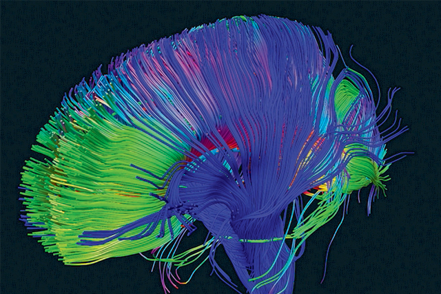
It is believed that the orientation of the space available in the animal due to the activity of so-called cell locations in the brain, which exhibit specific electrical activation in finding a specific point in the animal space. First, this activity exists in a wide spatial range. But after a short time after a new animal to enter the cell environment places that are in the hippocampus, a small area of the brain begin to demonstrate specific activity in certain areas of space and do not show it in the other.
If the animal appears several times in the same environment, there is a setting space cell activity when the animal is to more accurately differentiate themselves space. Especially if you find in some places it has something to do for an animal with aggressive behavior, electric shock, or some positive reinforcement, such as if the animal finds food. For example, the animal prefers the northern corner of the cell, where one neuron of the hippocampus will produce electrical activity, and the other is silent. And only when the animal starts to run across, say, in the southwest corner of the cage, another neuron will show other activity. There is a spatial differentiation of cells. Activity "bound" to the position of the animal cells in the space. The more mature neurons are, the more accurate differentiation occurs.
There is reason to believe that similar processes occur in the human brain. If we consider that the hippocampus performs a very similar function with the functions of the hippocampus to other mammals and one of his tasks - orientation in space, then in humans, this process should take place in a similar way.
A characteristic feature of the dentate gyrus of the hippocampus is continuing into adulthood neurogenesis - the generation of new neurons from stem cells. However, the role of these neurons adults behavior remained unknown until recently.
To improve brain function they recommend to use Cogitum, Nootropil, Cerebrolysin, Semax and Phenotropil.
Visualization of calcium signals allows scientists to monitor the electrical activity of neurons. The appearance of calcium in cells is closely associated with the generation of its actions or potentials bursts. There are special protein molecules, with calcium binding with which they begin to glow. This signal can be seen with a microscope, not penetrating into the brain tissue electrodes.
Optogenetics allows you to control the activity of individual neurons, stimulating or inhibiting their activity with light of a specific wavelength. This approach has expanded the arsenal of brain activity management. It is based on the introduction of light-sensitive cells in the membrane molecules channels which when illuminated can pass in one direction or another, into the cell or out of certain ions. The movement of the ions determines the cell's ability to produce an electrical discharge, or, on the contrary, break off. Lighting "activating" proteins lead to neuronal excitation, and the lighting "inhibiting", respectively, for inhibition. Thus, light can adjust optogenetics activity of individual neurons in restricted brain areas, without affecting adjacent structures.
By registering the calcium signal, the authors were able to establish that adults neurons were more active, demonstrating a greater incidence of calcium responses than their mature counterparts. This "tuning" activity of the cells is also dependent on the degree of maturity: unlike mature neurons, adults cells were not exactly spatially configured, which was expressed by activation of the same cells in the mouse being in different places of the virtual maze.
Then, we compared the variation of the activity of calcium in the brain in mice in sequential contact parts ( "A-B") or identical ( "B-B") maze. Different contexts differ in the olfactory, audible, tactile and visual environment. Mature neurons showed a high specificity for the activation position of the animal in a given point of the space, and their re-activation reproduced when placed in the same environment ( �B-B�). In contrast, the neurons did not show adults-playing activity at room successively animal in the same maze.
The final question was about the involvement adults neurons in the recognition of the new situation. Authors trained mice, combining the space into a new environment with an electric shock. The acquisition of such experience leads to a modification of animal behavior during the second hit in the same situation. Using optogenetic construct for inactivation light immature neurons of the hippocampus dentate gyrus in teaching, Danielson and his colleagues found that the activity of these cells is necessary for the acquisition of the memory of the dangers of the new situation.
Finally, the inactivation of neurons adult dentate gyrus of mice in the "A" camera, where they received an electric shock, do not prejudice the ability to distinguish similar, but the security camera "B". However, if the inactivation was performed at finding a mouse in the "B" chamber, it is difficult to secure the distinction between hazardous and contexts.
Thus, the authors were able to establish some important facts. Firstly, in the new situation adult immature neurons exhibit a higher level of excitability than their mature counterparts. However, they are less specific in space representation. Second, the need to set again appeared to discriminate neurons similar environments in vivo. The authors suggest that, ripening, adult neurons acquire the ability to more accurately encode space and more accurately distinguish between similar situation.